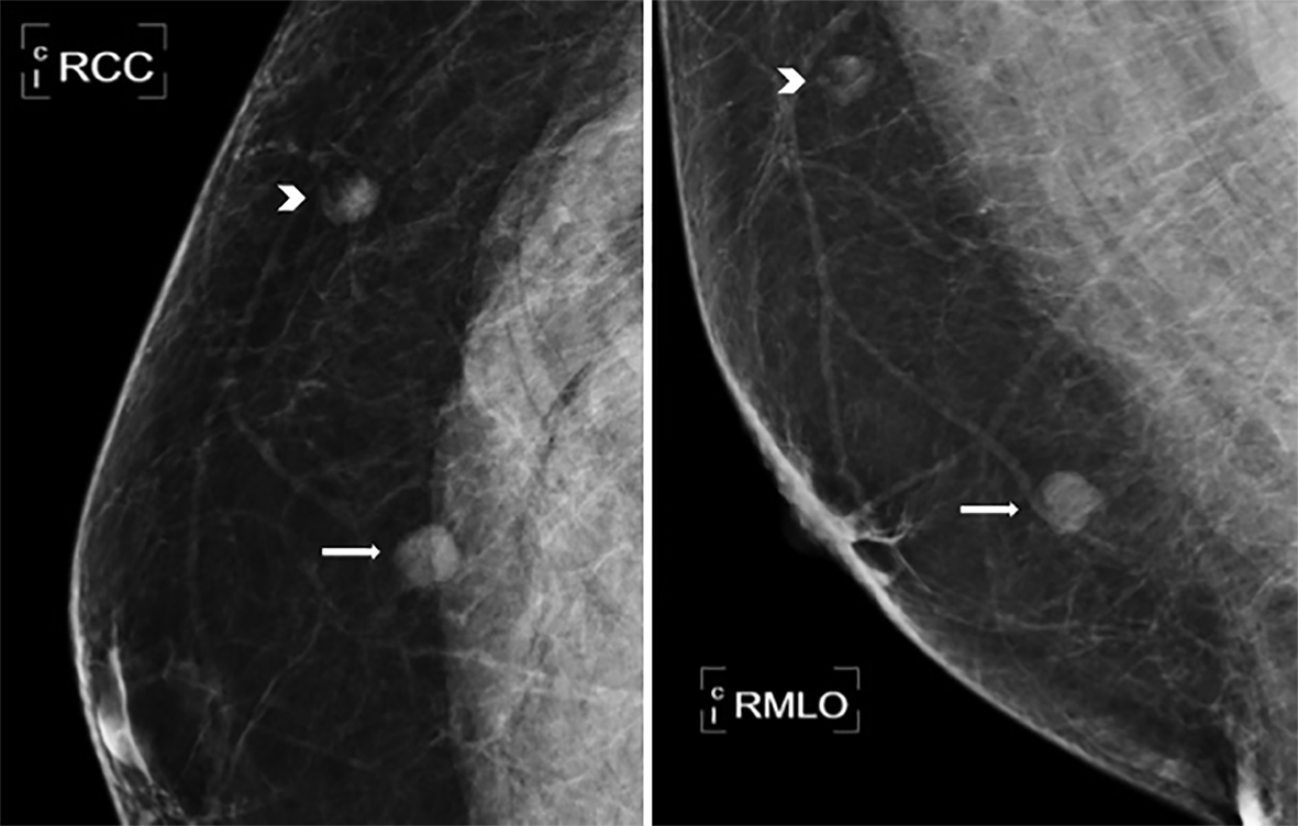
Figure 1. Mammogram of two well-defined lesions seen at 08:00 (arrow) and 11:00 (arrowhead).
| Journal of Medical Cases, ISSN 1923-4155 print, 1923-4163 online, Open Access |
| Article copyright, the authors; Journal compilation copyright, J Med Cases and Elmer Press Inc |
| Journal website http://www.journalmc.org |
Case Report
Volume 7, Number 8, August 2016, pages 323-325
Hemangioma in the Male Breast
Figures