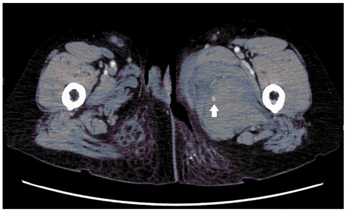
Figure 1. Contrast-enhanced CT image of the patient’s left thigh, showing a pseudoaneurysm (arrow) and hematoma in the adductor compartment.
| Journal of Medical Cases, ISSN 1923-4155 print, 1923-4163 online, Open Access |
| Article copyright, the authors; Journal compilation copyright, J Med Cases and Elmer Press Inc |
| Journal website http://www.journalmc.org |
Case Report
Volume 7, Number 7, July 2016, pages 299-302
Spontaneous Rupture of a Deep Femoral Pseudoaneurysm Mimicking Lymphedema After Radical Hysterectomy in a Woman Who Was Receiving Warfarin
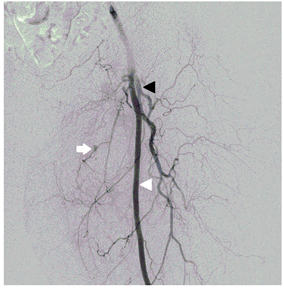
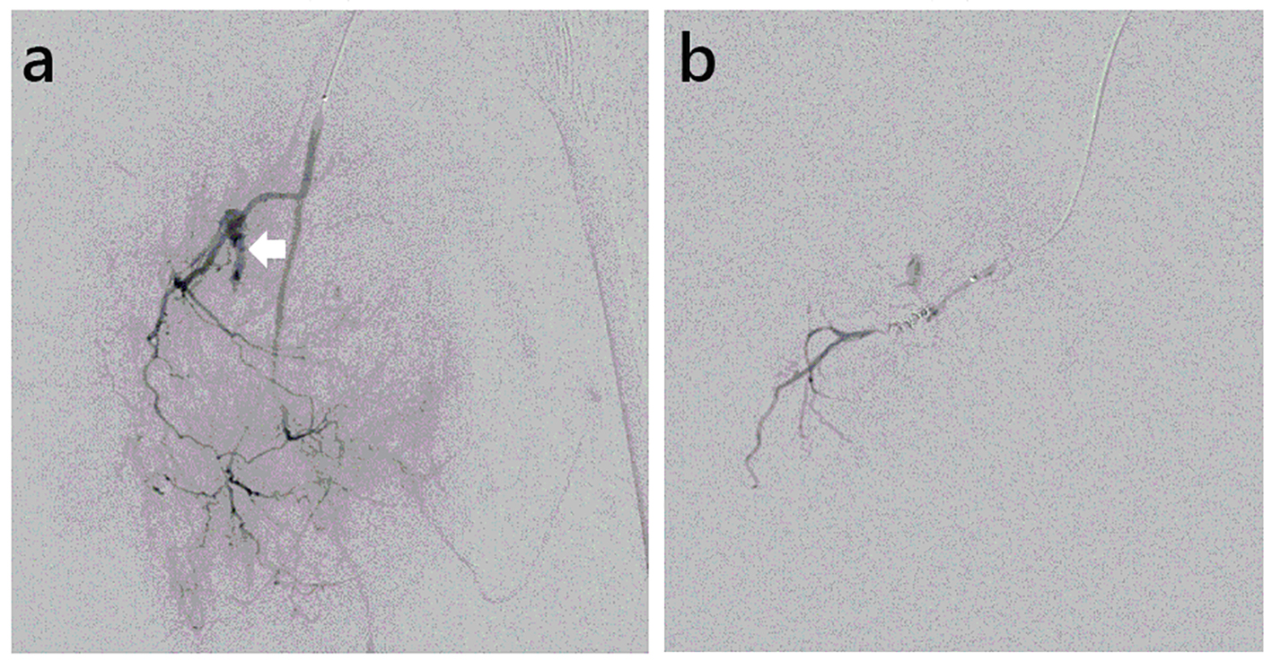
Figures