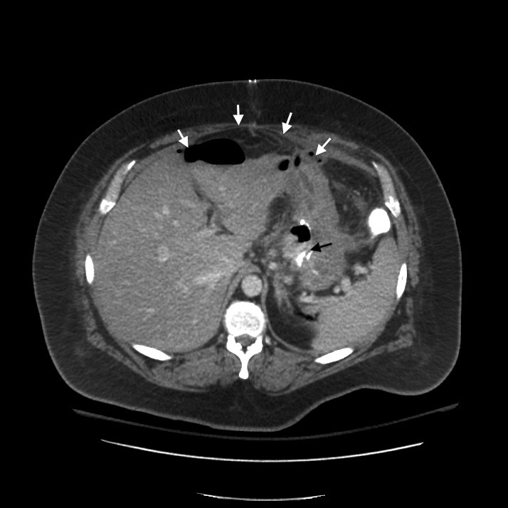
Figure 1. Computed tomography of abdomen at admission, demonstrating a large rim-enhancing multiloculated air and fluid collection (white arrows) near the gastrojejunal anastomosis (black arrow).
| Journal of Medical Cases, ISSN 1923-4155 print, 1923-4163 online, Open Access |
| Article copyright, the authors; Journal compilation copyright, J Med Cases and Elmer Press Inc |
| Journal website http://www.journalmc.org |
Case Report
Volume 2, Number 5, October 2011, pages 194-196
Successful Endoscopic Management of Gastrojejunal Anastomotic Leak Following Open Roux-en-Y Gastric Bypass
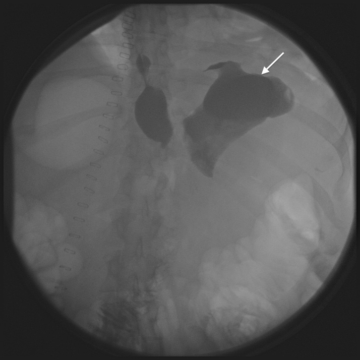
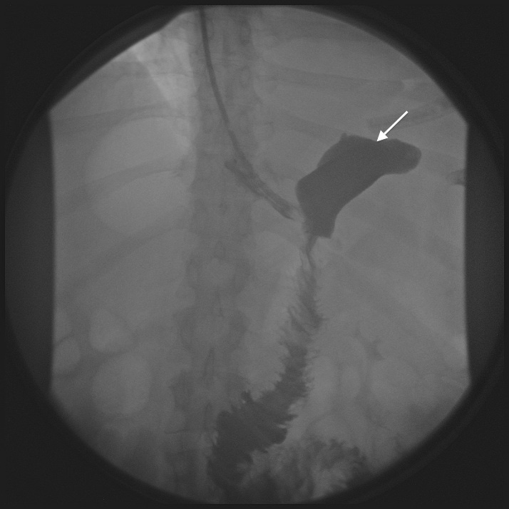
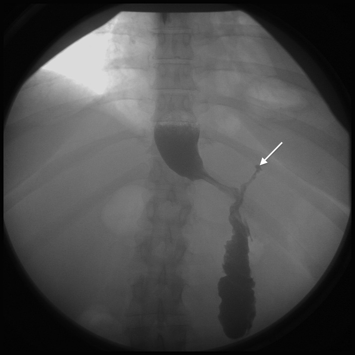
Figures