Journal of Medical Cases, ISSN 1923-4155 print, 1923-4163 online, Open Access
Article copyright, the authors; Journal compilation copyright, J Med Cases and Elmer Press Inc
Journal website http://www.journalmc.org

Case Report

Volume 7, Number 6, June 2016, pages 216-219


A Reminder of Treatments Past

Figures

Figure 1.
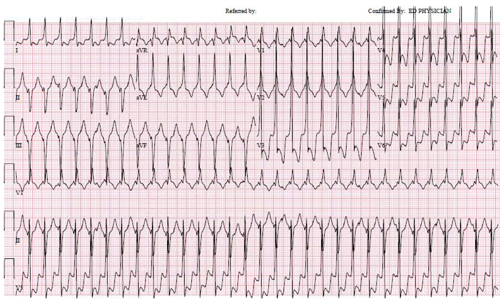
Figure 1. Regular wide complex tachycardia at 180 bpm, with a relatively narrow QRS duration of 120 bpm. The RBBB pattern with left axis deviation is typical for a fascicular VT originating from the LV septum.
Figure 2.
Figure 2. (A) Proximal circumflex artery occlusion with microchannels and left-to-left collateral circulation. (B) Close up view of A. (C) RAO view of left ventriculogram with an infero-basal aneurysm. (D) LAO view of left ventriculogram with an infero-basal aneurysm.
Figure 3.
Figure 3. 3D electroanatomic voltage map of the endocardial surface of the left ventricle, posterior view. Discrete posterior wall scar (bipolar, < 0.5 mV; region of red) extends from the base to mid ventricle.
Figure 4.
Figure 4. 3D electroanatomic map of the left ventricular septum, RAO caudal view. The sites of Purkinje potentials are marked (light purple dots) and outline the course of the branches of the left posterior fascicle.