Journal of Medical Cases, ISSN 1923-4155 print, 1923-4163 online, Open Access
Article copyright, the authors; Journal compilation copyright, J Med Cases and Elmer Press Inc
Journal website http://www.journalmc.org

Case Report

Volume 7, Number 8, August 2016, pages 315-319


Thymoma Presenting as Right-Side Heart Failure in a Young Healthy Patient: A Case Report and Literature Review

Figures

Figure 1.
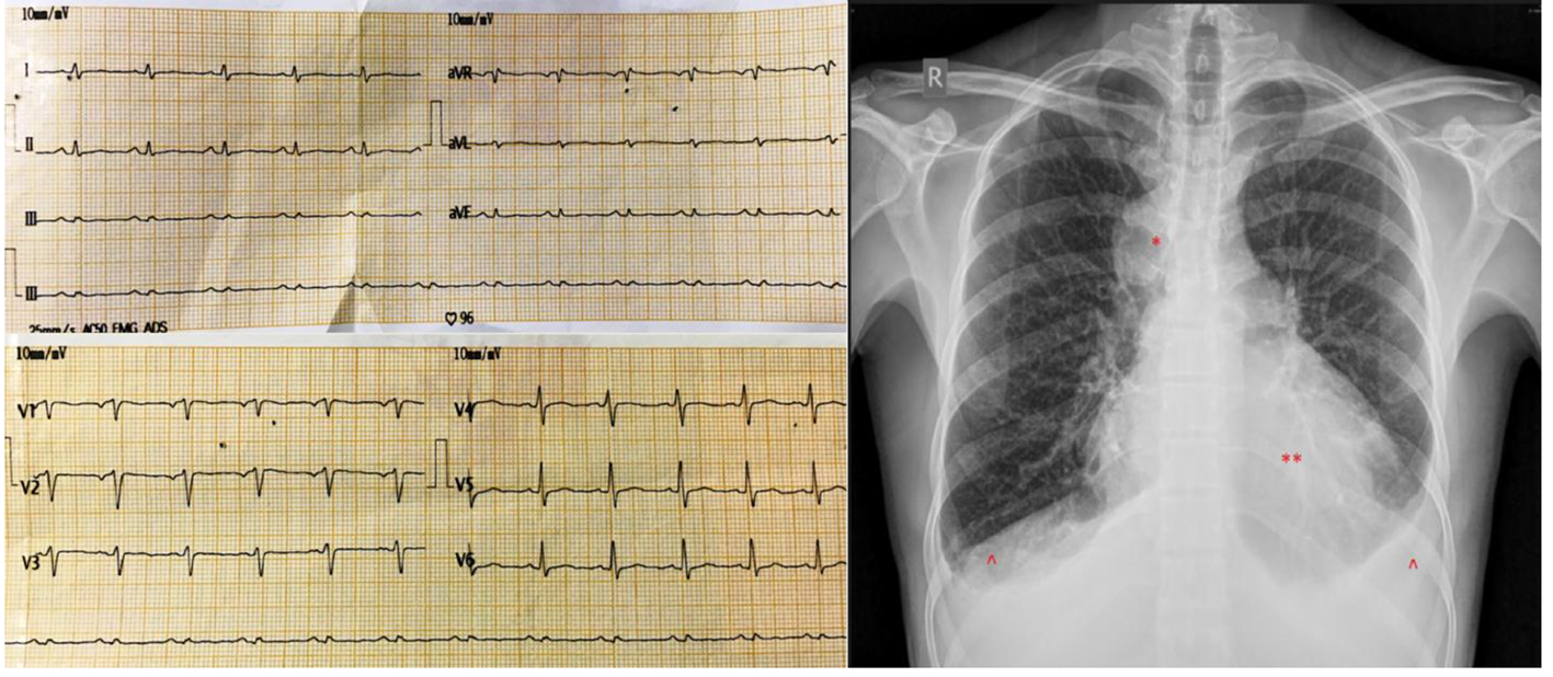
Figure 1. (Left) Electrocardiogram upon presentation showing microvoltage with sinus tachycardia. (Right) Chest roentgenogram upon presentation showing cardiomegaly (**) with bilateral pleural effusions (^) and a right upper paramediastinal round opacity (*).
Figure 2.
Figure 2. Echocardiogram showing a swinging heart in a massive circumferential pericardial effusion. LV: left ventricle; RV: right ventricle; LA: left atrium; RA: right atrium; Ao: aorta; PE: pericardial effusion.
Figure 3.
Figure 3. Computed tomography scan of the chest showing a retrosternal mass (*) with massive pericardial effusion (PE) and bilateral pleural effusions (P).

Tables

Table 1. WHO Histologic Typing of Tumors of the Thymus
 
TypeCharacteristicsFrequency
Type AMedullary thymomaSpindle cells; medullary9%
Type ABMixed thymomaMixed cellularity24%
Type B1Predominantly cortical thymomaLymphocyte-rich13%
Type B2Cortical thymomaPredominantly lymphocytic24%
Type B3Well differentiated thymic carcinomaEpithelial cells with mild atypia15%
Type CThymic carcinomaHighly atypical cells15%


Table 2. Masaoka Staging System for Thymoma and Thymic Carcinoma
 
StageCharacteristic5-year overall survival
Stage IMacroscopically and microscopically completely encapsulated94-100%
Stage IIAMicroscopic transcapsular invasion86-95%
Stage IIBMacroscopic invasion into surrounding fatty tissue or grossly adherent to but not through the mediastinal pleura or pericardium86-95%
Stage IIIAMacroscopic invasion into pericardium or lung without great vessel invasion56-69%
Stage IIIBMacroscopic invasion into pericardium or lung with great vessel invasion56-69%
Stage IVAPleural or pericardial dissemination11-50%
Stage IVBLymphogenous or hematogenous metastases11-50%