Figures
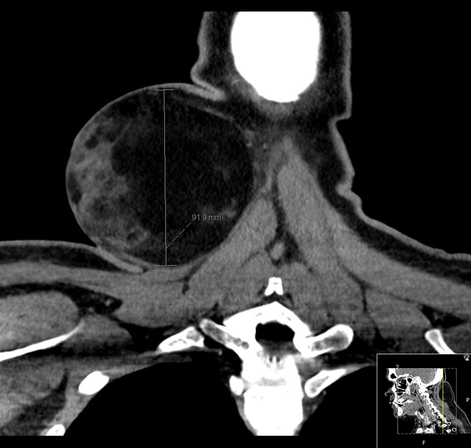
Figure 1. Coronal CT of a large right posterior neck triangle mass abutting the sternocleidomastoid muscle.
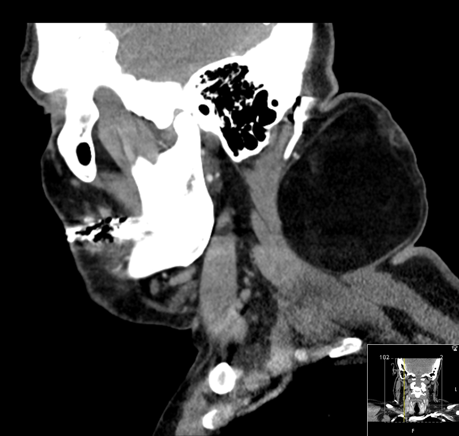
Figure 2. Sagittal CT image delineating the dimensions of the tumor.
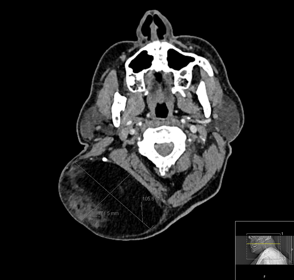
Figure 3. Axial CT image delineating the dimensions of the tumor.
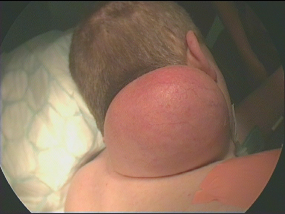
Figure 4. Pre-operative photograph of a large right posterior neck mass.
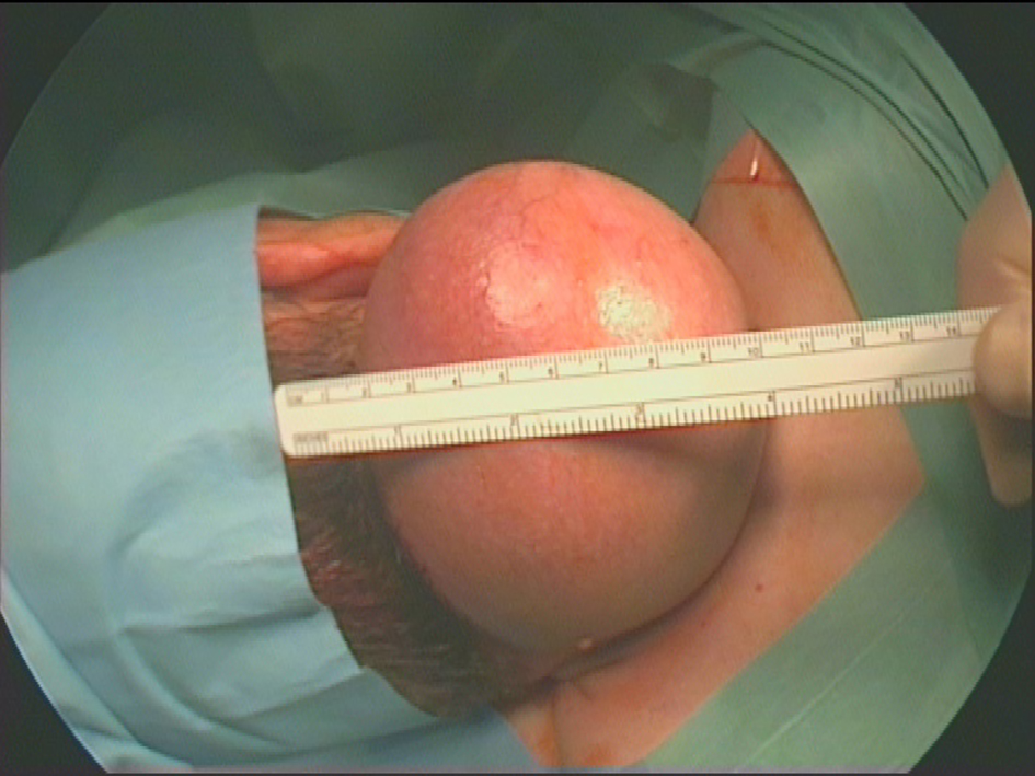
Figure 5. Pre-operative photograph of a large right posterior neck mass (draped).
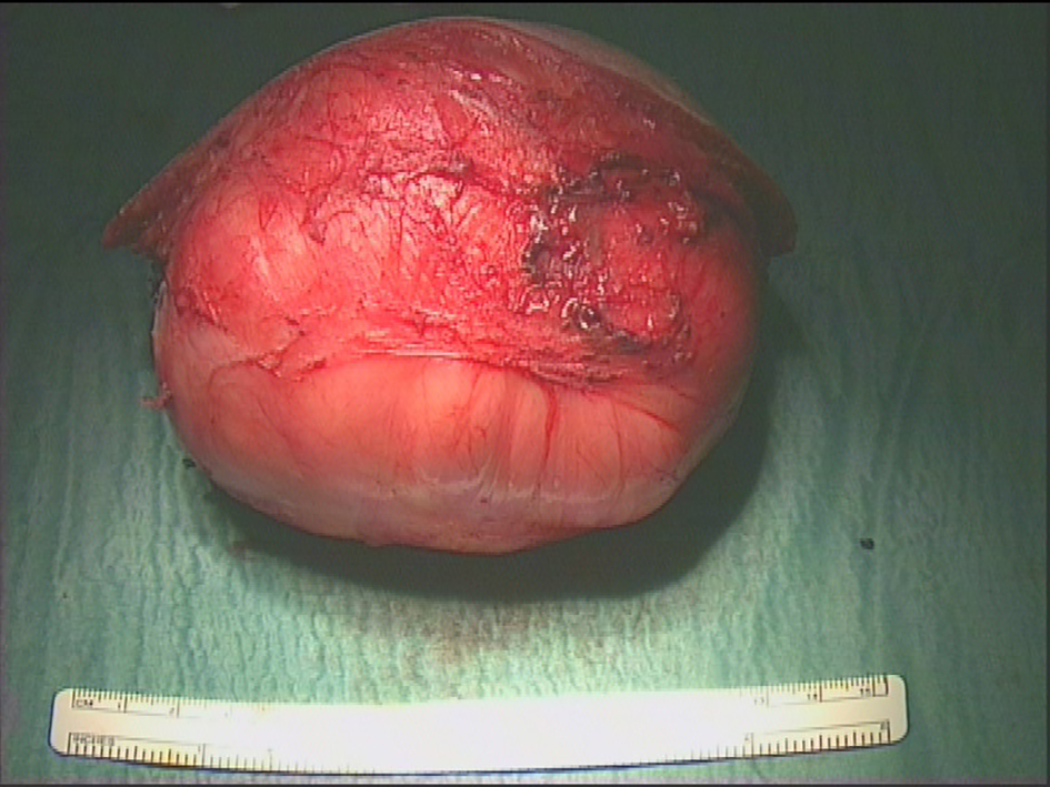
Figure 6. Lesion following complete resection illustrating intact capsule.
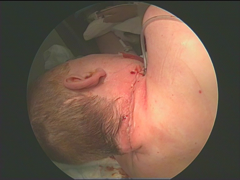
Figure 7. Post-operative appearances following closure.
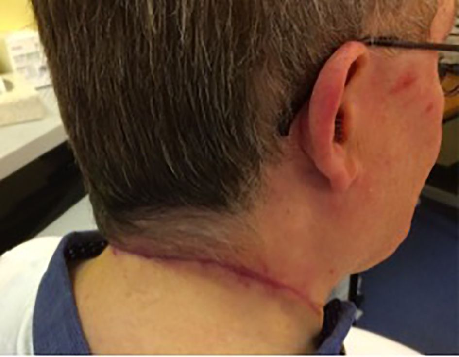
Figure 8. Appearance of the wound 2 months post-operatively.
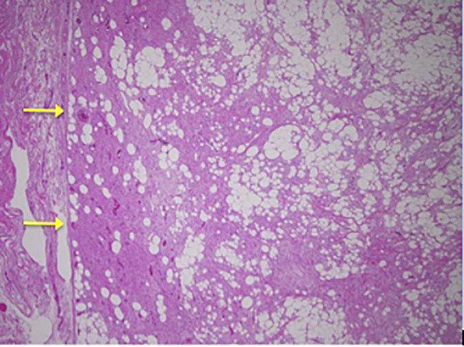
Figure 9. Low power image showing capsule of lesion on left.
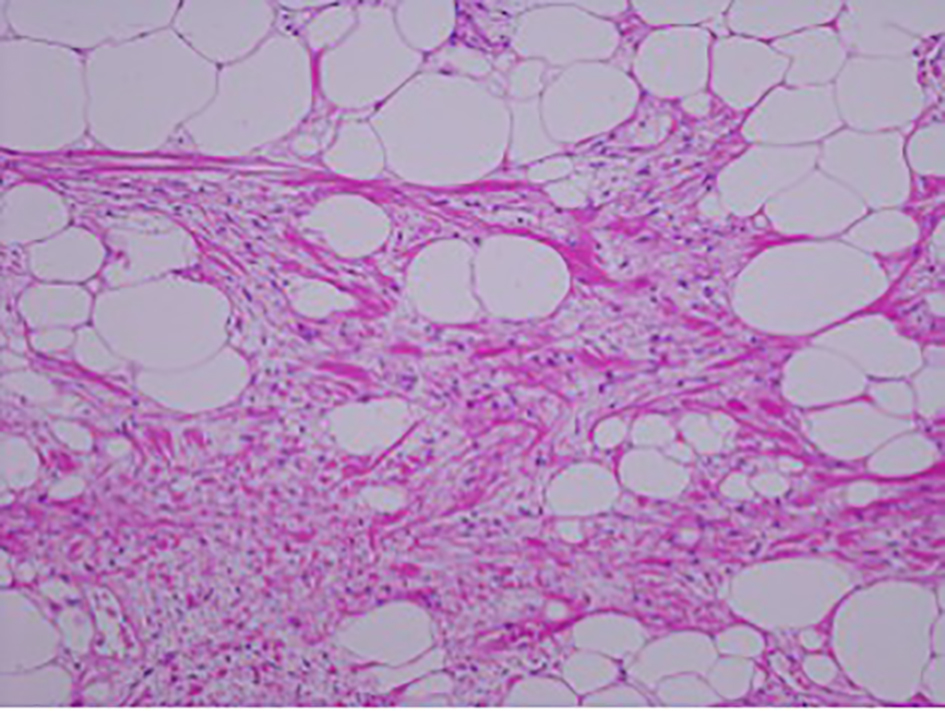
Figure 10. Magnification image (× 10) showing spindle cells admixed with ropey collagen and mature adipocytes in a myxoid matrix.
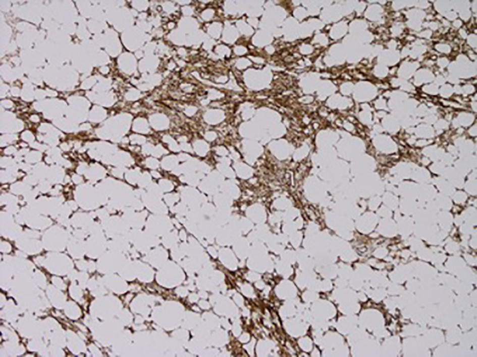
Figure 11. CD34 immunostain showing positivity in the spindle cell areas (brown coloration).