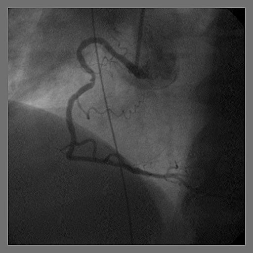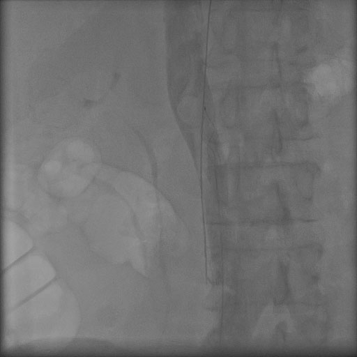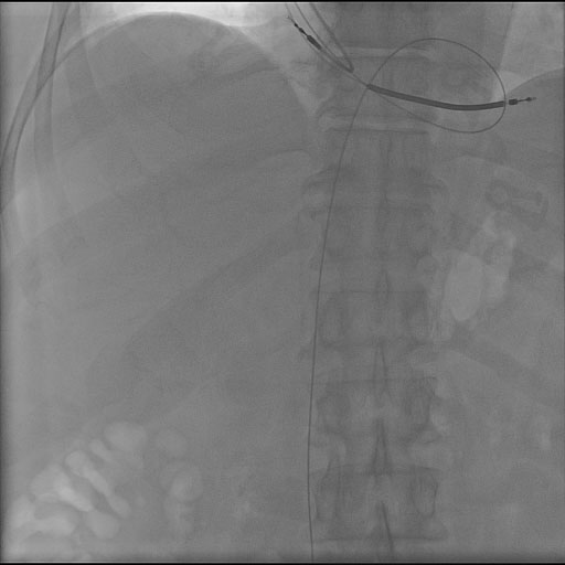
Figure 1. J-wire was noted to run from the superior vena cava (SVC) to the right atrium, looped through the right ventricle and into the inferior vena cava (IVC) under fluoroscopy.
| Journal of Medical Cases, ISSN 1923-4155 print, 1923-4163 online, Open Access |
| Article copyright, the authors; Journal compilation copyright, J Med Cases and Elmer Press Inc |
| Journal website http://www.journalmc.org |
Case Report
Volume 2, Number 4, August 2011, pages 159-161
A Novel Approach to the Removal of a Retained Guidewire
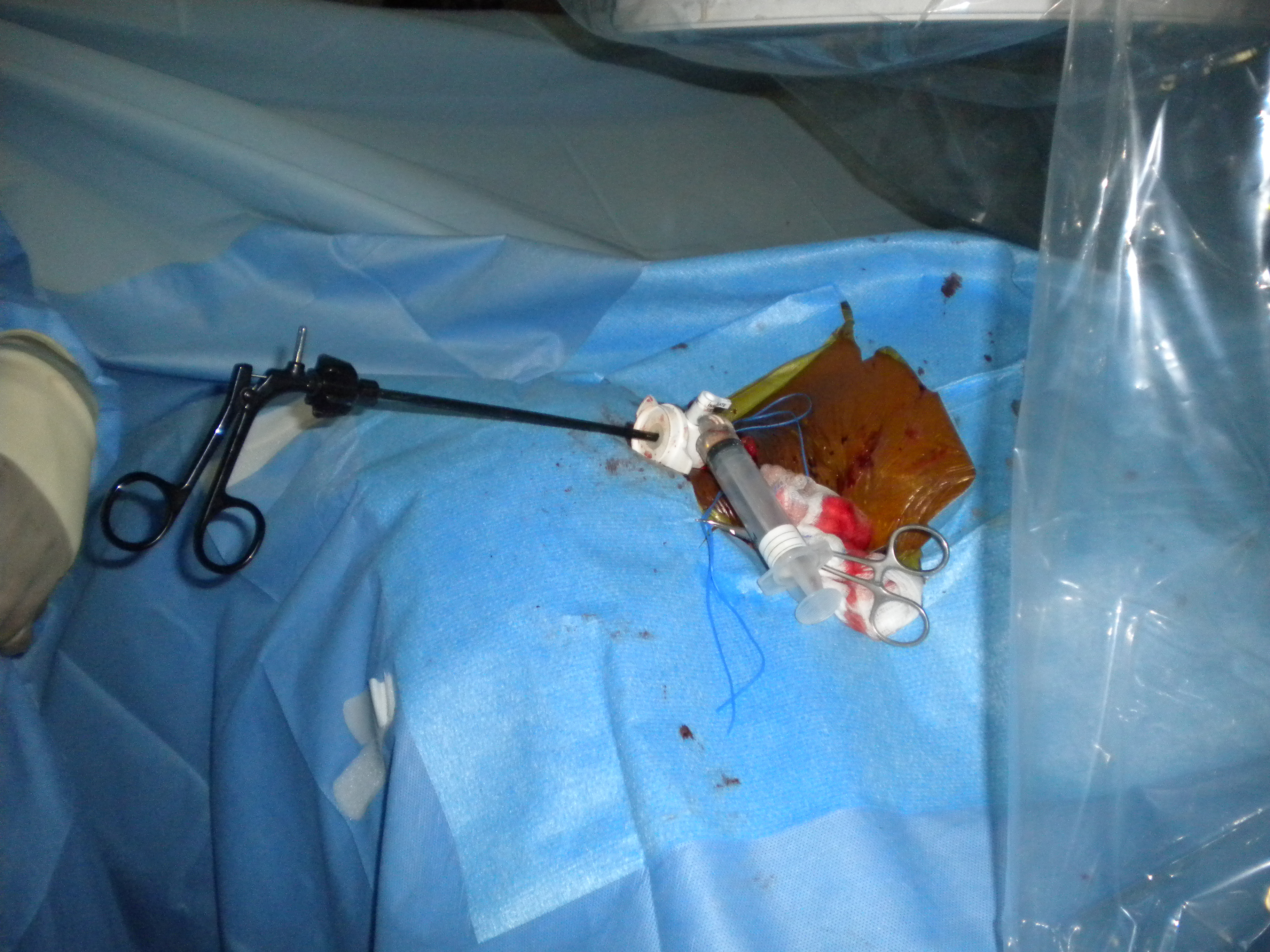
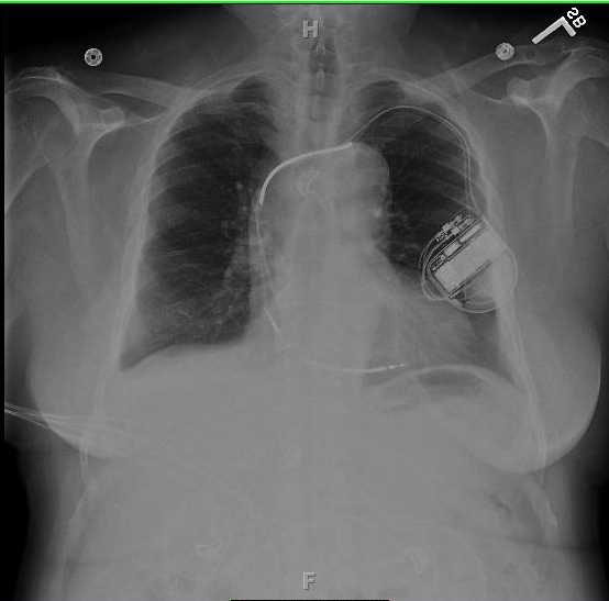
Figures