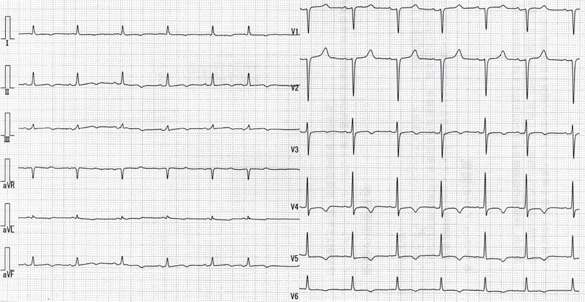
Figure 1. Electrocardiography shows normal sinus rhythm with inverted T in I, II, III, aVL, aVf, V3-6 leads.
| Journal of Medical Cases, ISSN 1923-4155 print, 1923-4163 online, Open Access |
| Article copyright, the authors; Journal compilation copyright, J Med Cases and Elmer Press Inc |
| Journal website http://www.journalmc.org |
Case Report
Volume 5, Number 9, September 2014, pages 498-501
Constrictive Pericarditis: Rapid Progression of Pericardial Calcification in a Patient With Hemodialysis and Coronary Artery Bypass Surgery
Figures
Table
| Pressure (mm Hg) | Pre | Post |
|---|---|---|
| RA: right atrium; RV: right ventricle; PA: pulmonary artery; PCW: pulmonary capillary wedge; LV: left ventricle; Ao: aorta; a: a wave; v: v wave; m: mean pressure; s: systolic pressure; d: diastomic pressure; ed: end diastolic pressure. The preoperative hemodynamic measurements showed near equalization of diastolic pressure in all chambers and an early dip and plateau were seen in the pressure tracings of both ventricles. Pressure data obtained from cardiac catheterization at 3 weeks after pericardiectomy revealed hemodynamical improvement. | ||
| RA (a/v/m) | 18/16/15 | 4/2/1 |
| RV (s/d/ed) | 59/12/20 | 36/0/4 |
| PA (s/d/m) | 56/23/24 | 46/15/26 |
| PCW (a/v/m) | 36/20/25 | 18/19/15 |
| LV (s/d/ed) | 147/11/22 | Not done |
| Ao (s/d/m) | 144/60/95 | Not done |