Journal of Medical Cases, ISSN 1923-4155 print, 1923-4163 online, Open Access
Article copyright, the authors; Journal compilation copyright, J Med Cases and Elmer Press Inc
Journal website http://www.journalmc.org

Case Report

Volume 5, Number 9, September 2014, pages 488-490


Veno-Venous Extracorporeal Membrane Oxygenation for Fat Embolism

Figures

Figure 1.
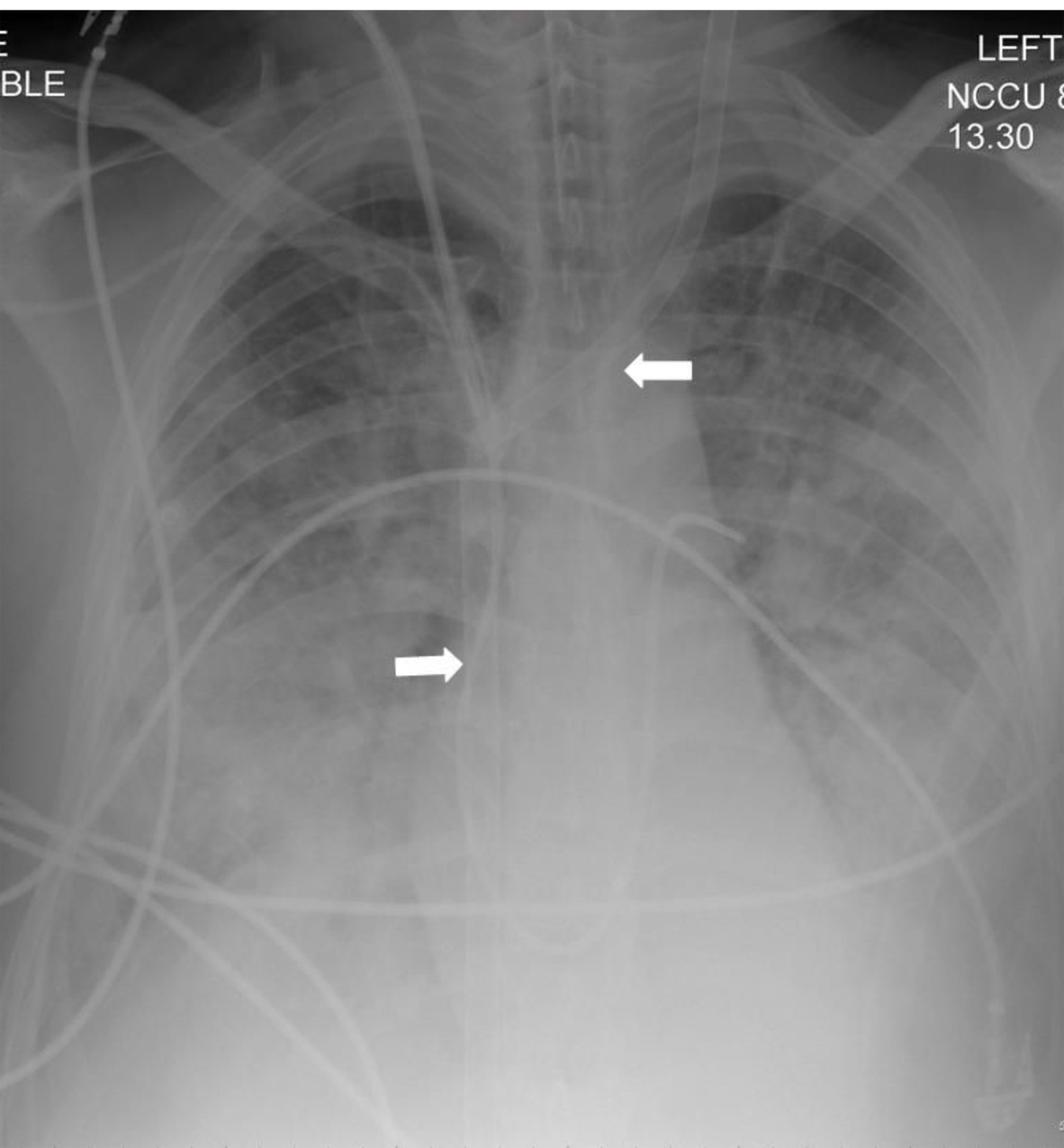
Figure 1. A rotated supine chest radiograph on admission demonstrates clear lungs and pleural spaces.
Figure 2.
Figure 2. Supine chest radiograph over the next 12 h showed progressive worsening in the bilateral alveolar opacification predominantly involving the mid- and lower zones and development of small to moderate pleural effusions. Note the presence of the veno-venous ECMO cannulae (block arrows). Despite the lack of recirculation of oxygenated blood the IVC cannula was withdrawn by 5 cm.
Figure 3.
Figure 3. Allowing for the rotation, the AP chest radiograph post decannulation demonstrates complete resolution of the parenchymal opacification and pleural effusions.
Figure 4.
Figure 4. Oxygenator output: x-axis: days on ECMO; y-axis: PO2 of blood post oxygenator.