Journal of Medical Cases, ISSN 1923-4155 print, 1923-4163 online, Open Access
Article copyright, the authors; Journal compilation copyright, J Med Cases and Elmer Press Inc
Journal website http://www.journalmc.org

Case Report

Volume 5, Number 9, September 2014, pages 511-514


An Atypical Presentation of Small Bowel Metastasis From a Previously Undiagnosed Primary Lung Adenocarcinoma

Figures

Figure 1.
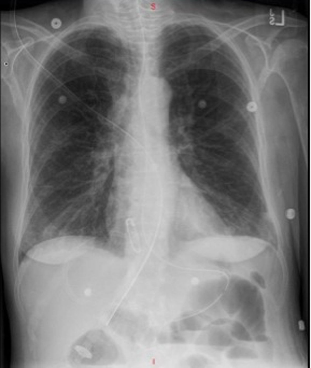
Figure 1. Posterior/anterior (PA) chest X-ray on day of admission showing no acute disease and normal pulmonary vascularity.
Figure 2.
Figure 2. CT of abdomen and pelvis without contrast showing multiple dilated loops of small bowel throughout the abdomen with compression of the loops comparable with a high-grade small bowel obstruction.
Figure 3.
Figure 3. (A) H&E stain of invasive carcinoma into the small bowel submucosa with atypical mitosis (arrow). (B) H&E stain of the small bowel demonstrating undifferentiated adenocarcinoma with glandular formation (arrow head).
Figure 4.
Figure 4. (A) Pericardial fluid aspiration showing glandular structures. (B) Tumor cells within the pericardial fluid aspirate. (C) Immunohistochemical stain positive for TTF-1 (lung and thyroid cancer marker). (D) Immunohistochemical stain positive for MOC-31 (adenocarcinoma marker).