Journal of Medical Cases, ISSN 1923-4155 print, 1923-4163 online, Open Access
Article copyright, the authors; Journal compilation copyright, J Med Cases and Elmer Press Inc
Journal website http://www.journalmc.org

Case Report

Volume 5, Number 8, August 2014, pages 463-465


Ileitis Is Not Always Crohn’s Disease: Case Report

Figures

Figure 1.
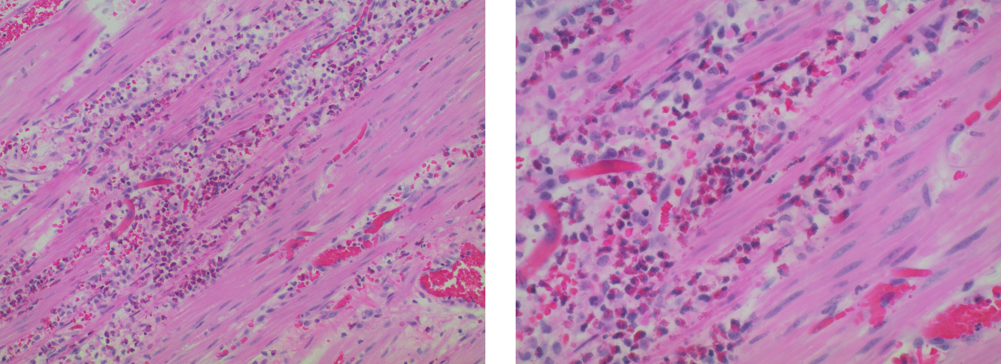
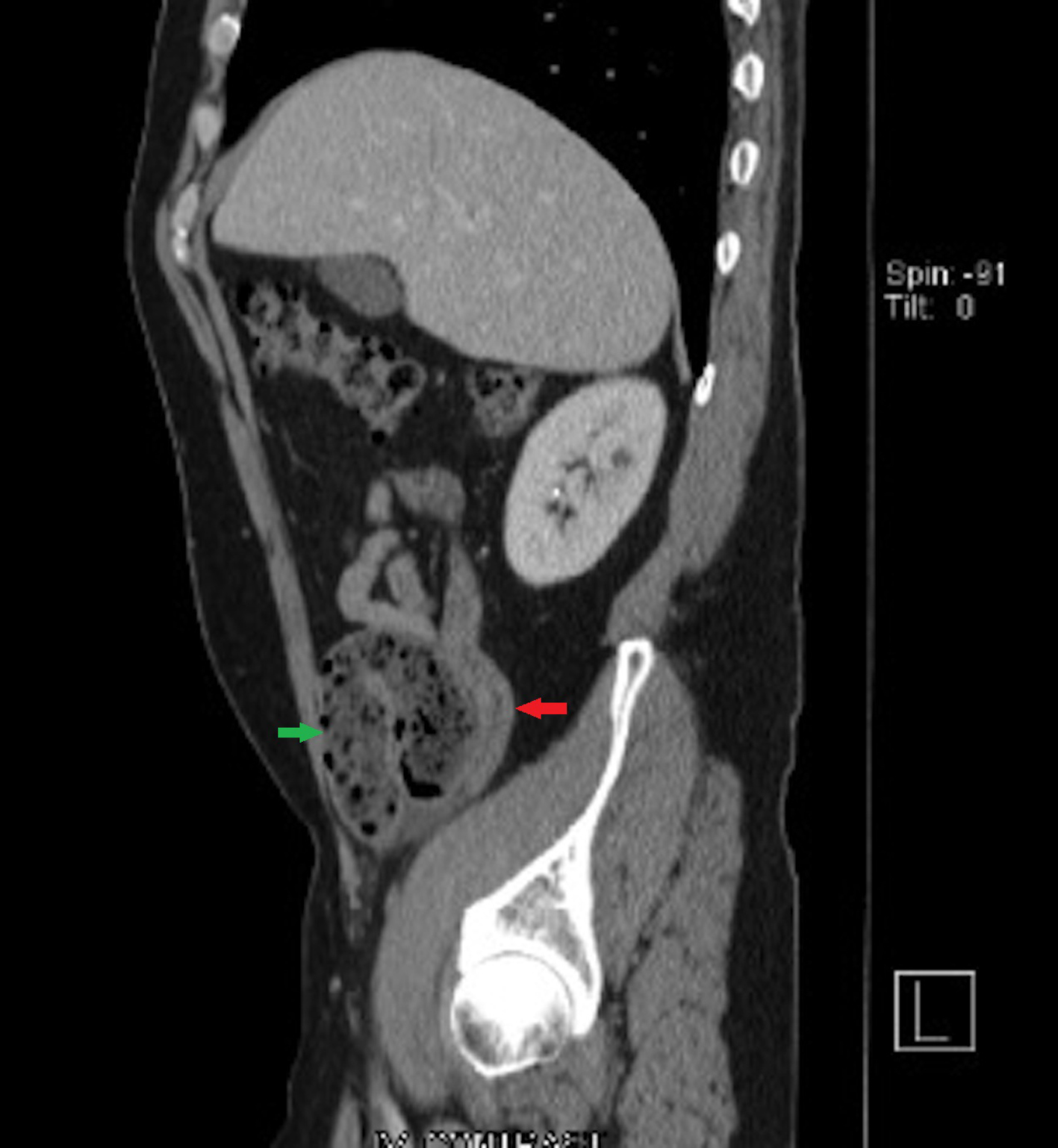
Figure 1. CT of abdomen and pelvis with intravenous contrast showed bowel wall thickening and inflammatory changes around the terminal ileum. Red arrow indicates bowel wall thickening and inflammatory changes around the terminal ileum (sagittal view). Green arrow indicates dilatation of the small bowel just proximal to ileitis area (sagittal view).
Figure 2.
Figure 2. Surgical pathology: dense infiltrate of eosinophils in the muscularis propria suggests EG.