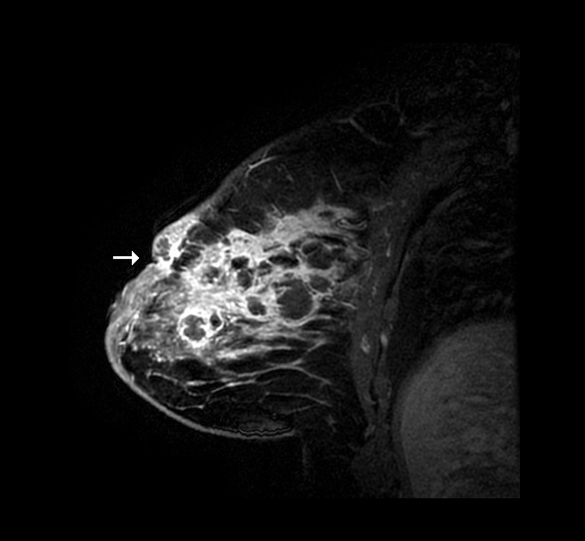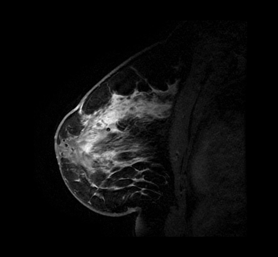
Figure 1. Contrast enhanced MRI sagittal view of the left breast demonstrating diffuse enhancement of the breast parenchyma.
| Journal of Medical Cases, ISSN 1923-4155 print, 1923-4163 online, Open Access |
| Article copyright, the authors; Journal compilation copyright, J Med Cases and Elmer Press Inc |
| Journal website http://www.journalmc.org |
Case Report
Volume 5, Number 8, August 2014, pages 430-434
Bilateral Idiopathic Granulomatous Mastitis: A Case of Noncaseating Granulomas in a Patient With Latent Tuberculosis
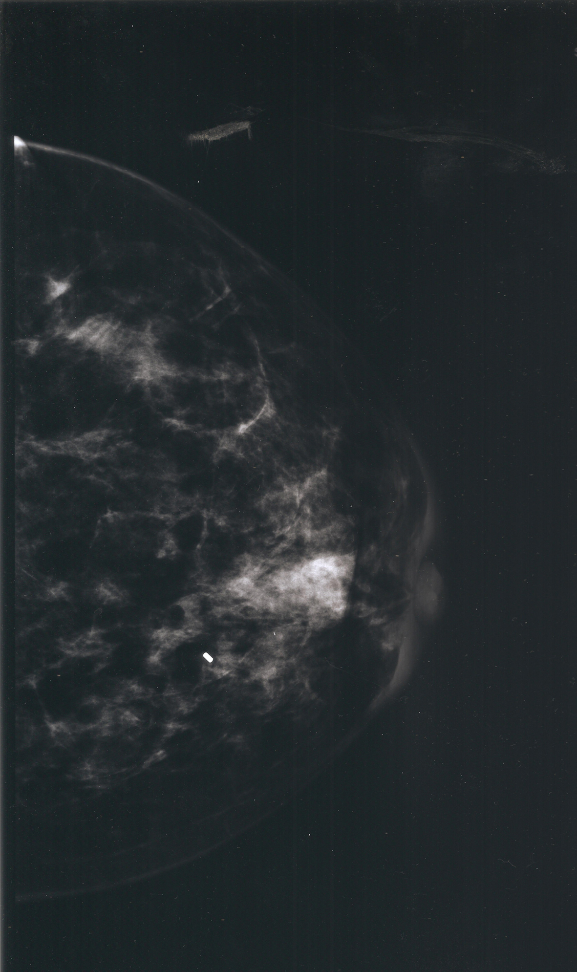
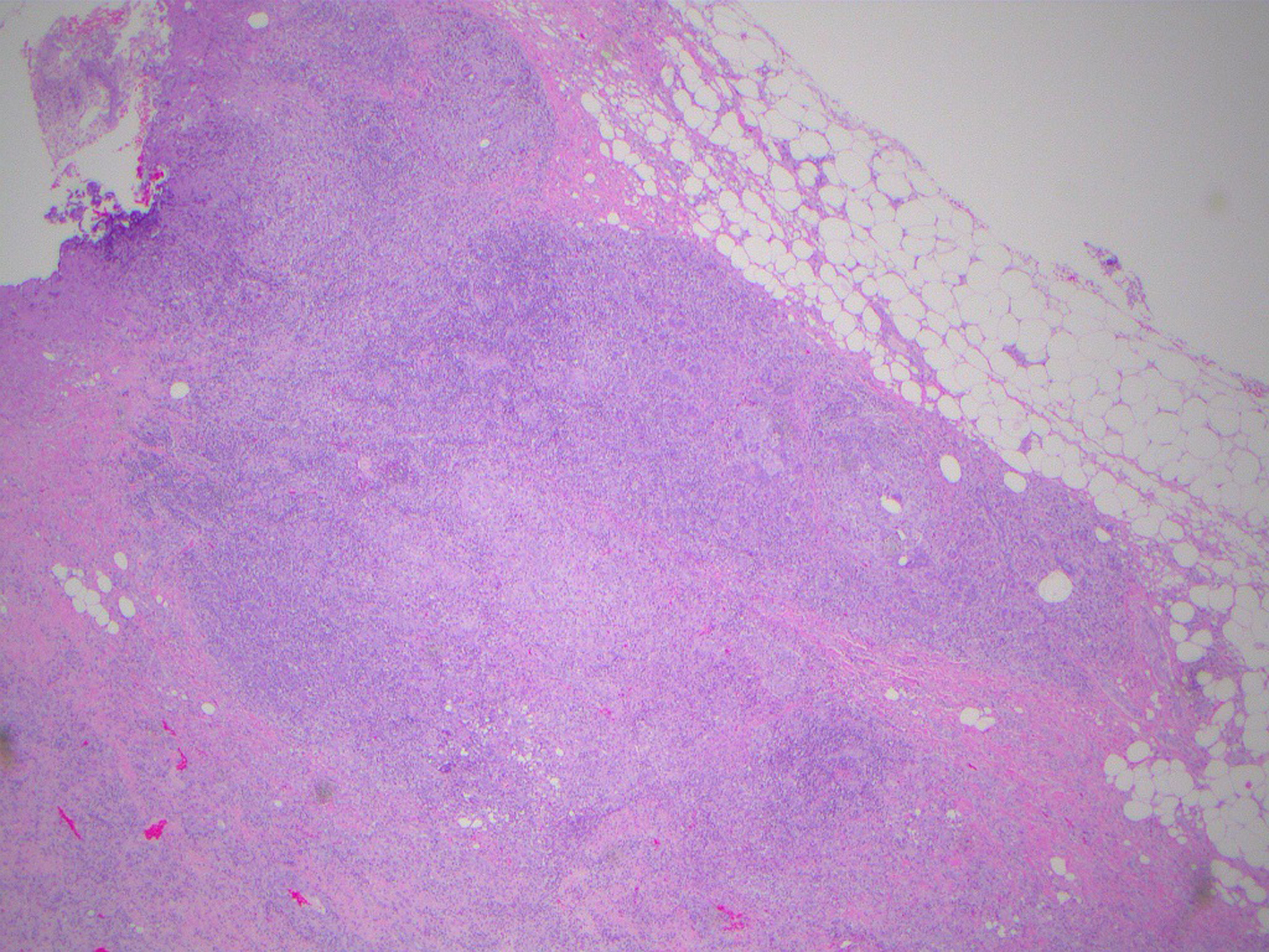
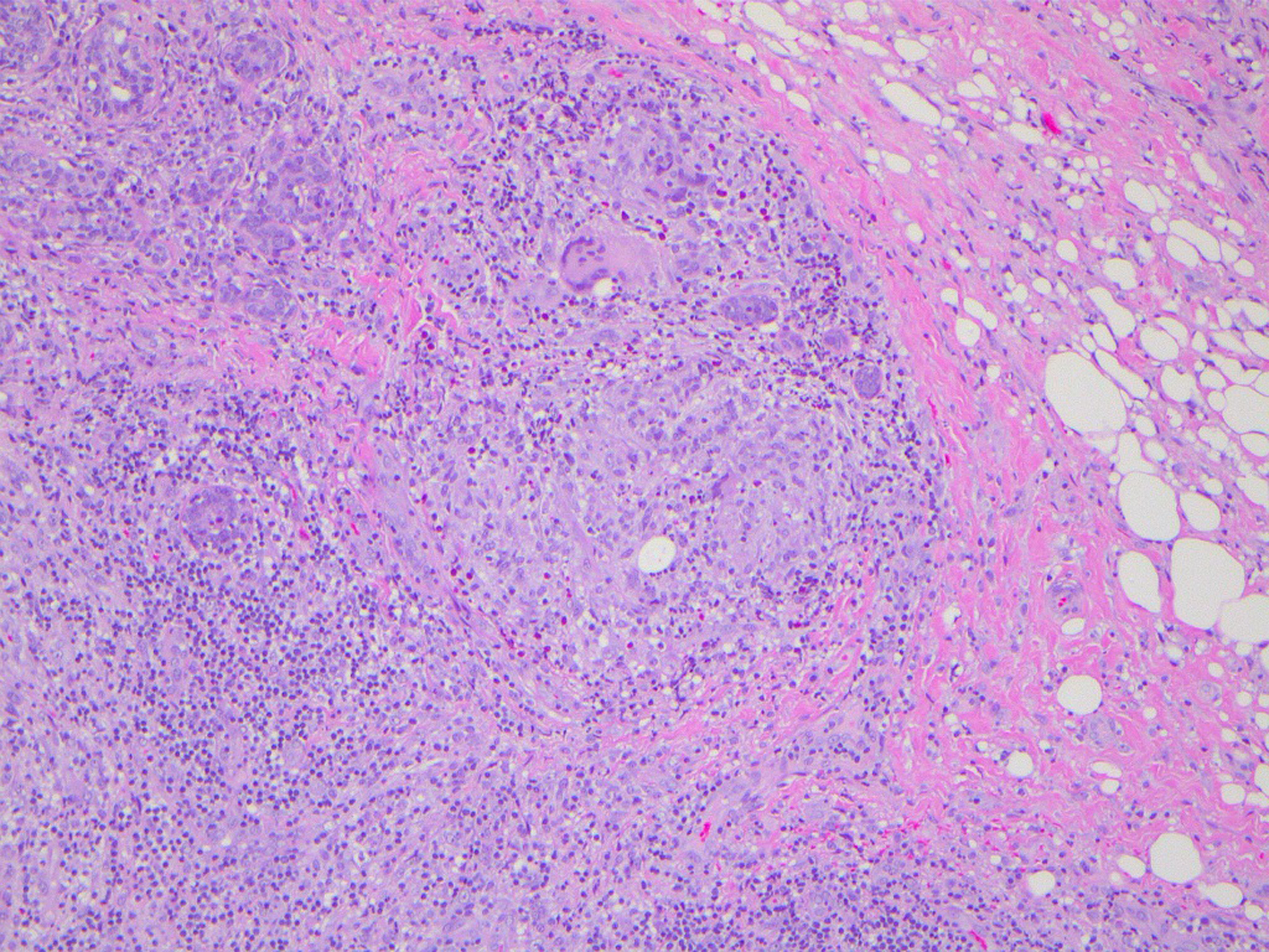
Figures