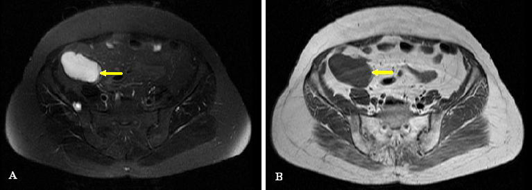
Figure 1. MRI of mass: (A) T2 sequence; (B) T1 sequence.
| Journal of Medical Cases, ISSN 1923-4155 print, 1923-4163 online, Open Access |
| Article copyright, the authors; Journal compilation copyright, J Med Cases and Elmer Press Inc |
| Journal website http://www.journalmc.org |
Case Report
Volume 5, Number 7, July 2014, pages 423-425
Laparoscopic Appendectomy for Giant Appendiceal Mucocele in an Elderly Patient: A Case Report
Figures