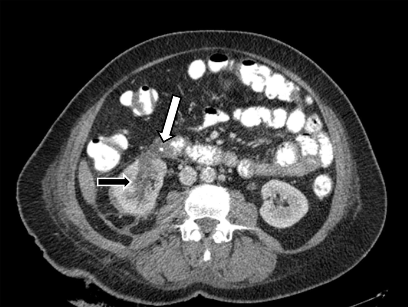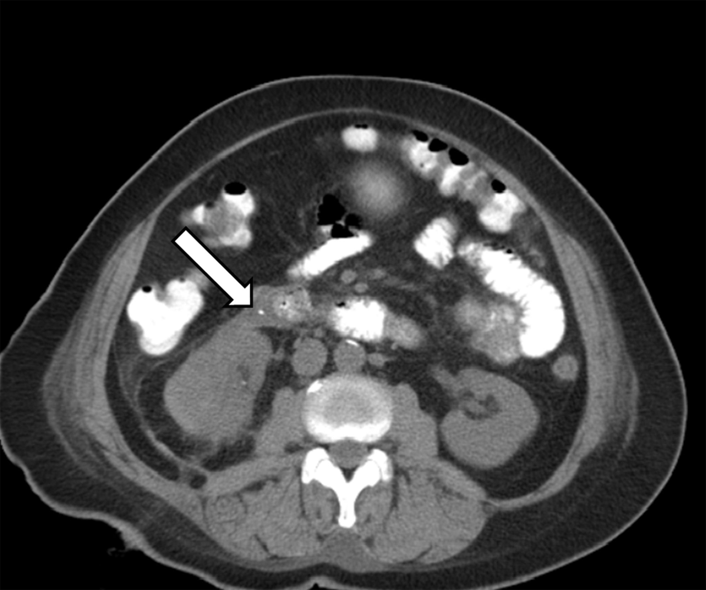
Figure 1. This CT image obtained right after the lithotripsy attempt shows a deformed right kidney together with a 3 mm opacity in the near vicinity of the duodenum (arrow), which was mistaken for extravasated oral contrast from the duodenum.
| Journal of Medical Cases, ISSN 1923-4155 print, 1923-4163 online, Open Access |
| Article copyright, the authors; Journal compilation copyright, J Med Cases and Elmer Press Inc |
| Journal website http://www.journalmc.org |
Case Report
Volume 5, Number 7, July 2014, pages 408-410
Iatrogenically Displaced Renal Calculus During Percutaneous Lithotripsy That Mimicked Duodenal Perforation by Resembling Leaked Oral Contrast
Figures