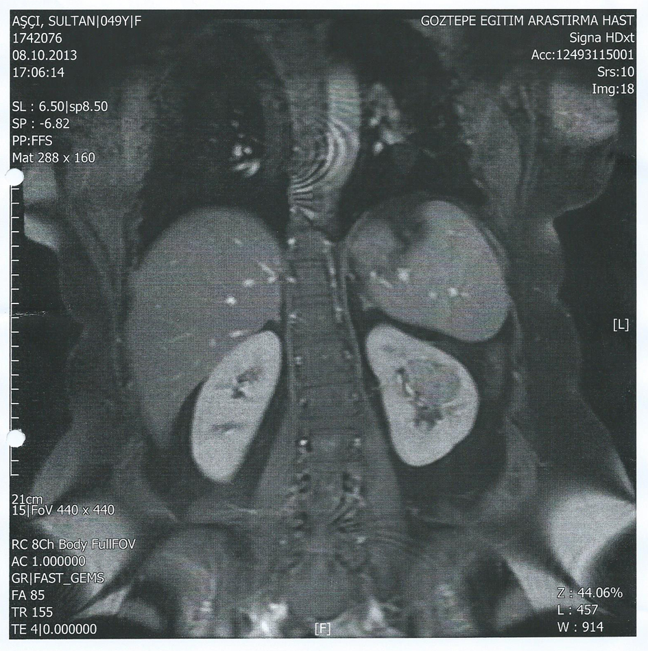
Figure 1. On coronal plane, on the contrasted, fat-saturated T1 images, 26 × 44 mm lobule contoured mass was seen.
| Journal of Medical Cases, ISSN 1923-4155 print, 1923-4163 online, Open Access |
| Article copyright, the authors; Journal compilation copyright, J Med Cases and Elmer Press Inc |
| Journal website http://www.journalmc.org |
Case Report
Volume 5, Number 6, June 2014, pages 362-365
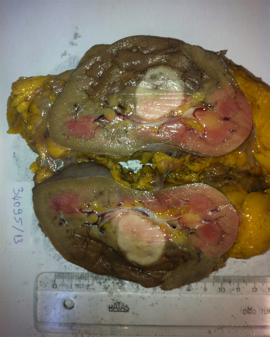
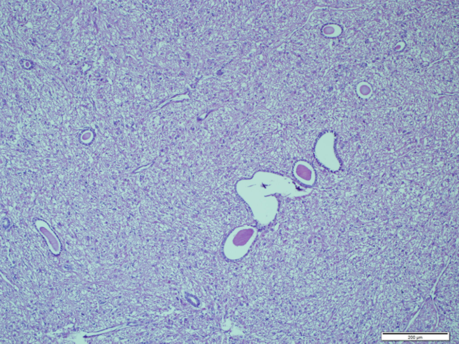
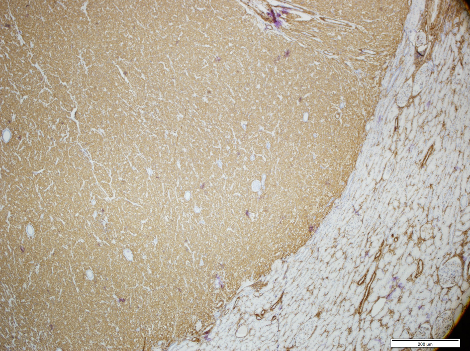
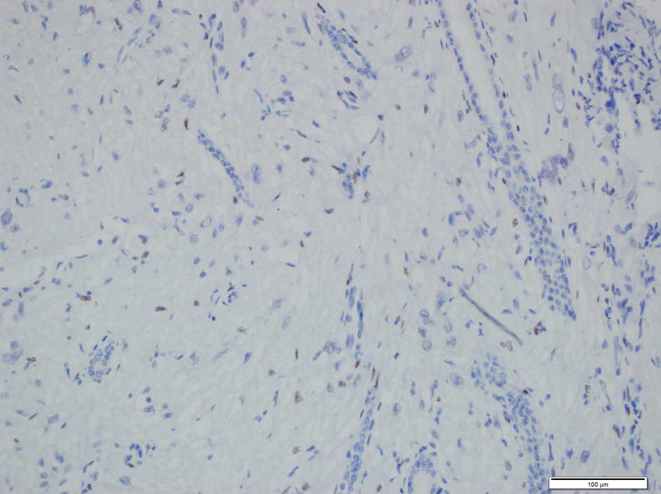
Mixed Epithelial and Stromal Tumor of the Kidney: A Rare Case Report
Figures