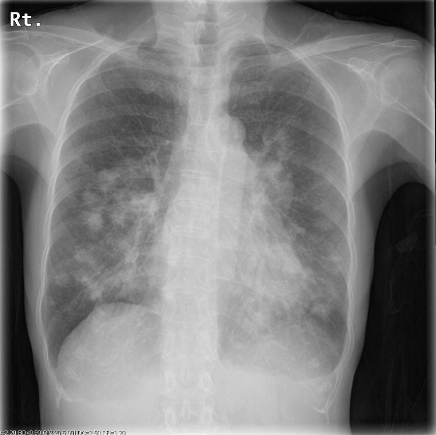
Figure 1. Chest radiograph on admission showing consolidation of the bilateral lungs.
| Journal of Medical Cases, ISSN 1923-4155 print, 1923-4163 online, Open Access |
| Article copyright, the authors; Journal compilation copyright, J Med Cases and Elmer Press Inc |
| Journal website http://www.journalmc.org |
Case Report
Volume 5, Number 5, May 2014, pages 279-282
Epstein-Barr Virus-Related Lymphoproliferative Disorder Caused by the Use of Antithymocyte Globulin and Cyclosporine for Aplastic Anemia
Figures
Table
| Hematology | Biochemistry | Serology | |||
|---|---|---|---|---|---|
| Abbreviations: sIL-2R, soluble interleukin-2 receptor. | |||||
| WBC | 13,100/µL | Alb | 3.0 g/dL | CRP | 2.78 mg/dL |
| Myelo | 1.0% | BUN | 33.1 g/dL | IgG | 756 mg/dL |
| Stab | 1.5% | Cr | 1.23 mg/dL | IgA | 307 mg/dL |
| Seg | 69.5% | T-Bil | 0.6 mg/dL | IgM | 89 mg/dL |
| Eosino | 0.5% | ALP | 312 U/L | Cyclosporine | 54.4 ng/mL |
| Ly | 17.0% | LDH | 272 U/L | sIL-2R | 6040 U/mL |
| Mono | 10.5% | AST | 17 U/L | ||
| RBC | 339 × 104/mL | ALT | 13 U/L | Arterial blood gas analysis (FiO2 0.24) | |
| Hb | 9.9 g/dL | γ-GTP | 83 U/L | pH | 7.450 |
| Hct | 29.0% | CK | 23 U/L | PaCO2 | 33.3 mmHg |
| Plt | 25.5 × 10⁴/mL | Ca | 8.8 mg/dL | PaO2 | 70.8 mmHg |
| Na | 135 mEq/L | HCO3- | 22.8 mEq/L | ||
| K | 4.3 mEq/L | Lactate | 1.0 mmol/L | ||
| Cl | 101 mEq/L | ||||
| Glu | 187 mg/dL | ||||
| β-D glucan | 5.7 pg/mL | ||||