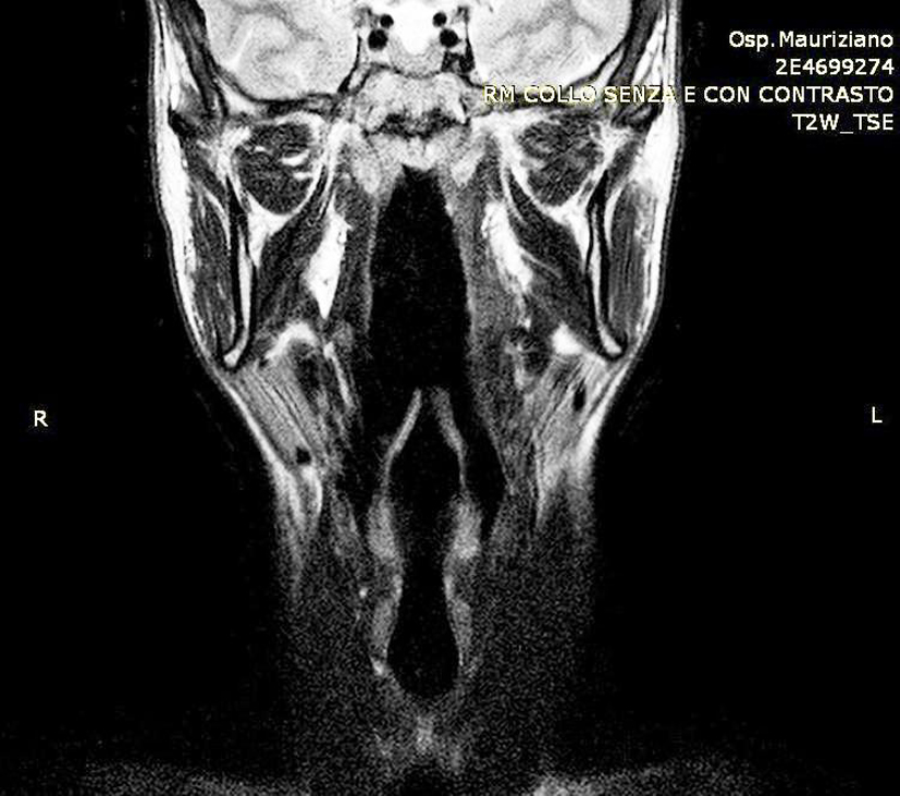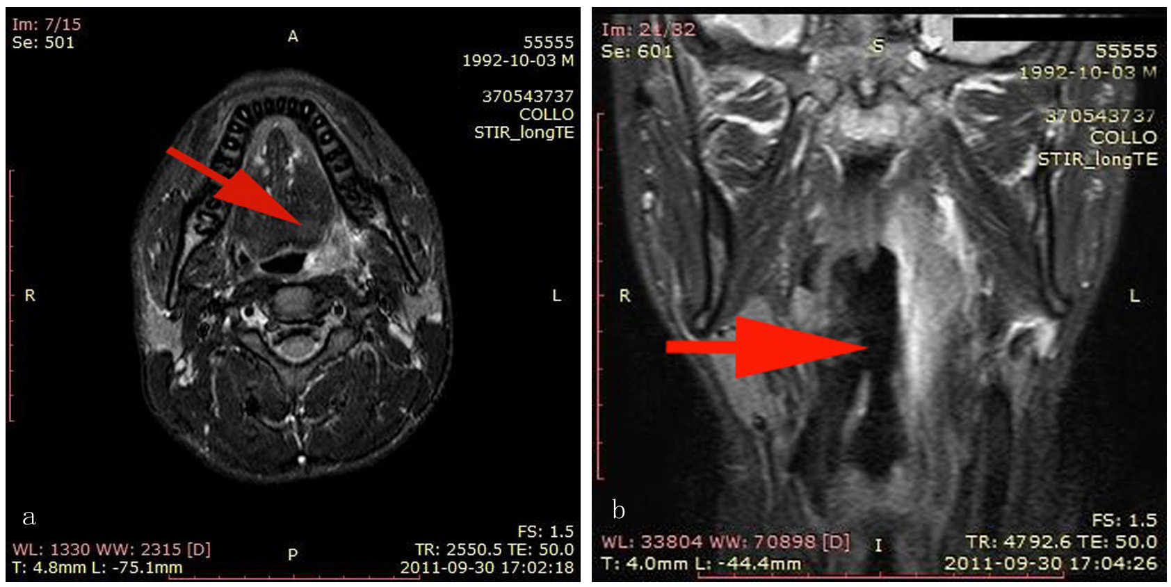
Figure 1. (a, b) MRI of the cervical region revealed a prestyloid parapharyngeal abscess on the left side (arrows).
| Journal of Medical Cases, ISSN 1923-4155 print, 1923-4163 online, Open Access |
| Article copyright, the authors; Journal compilation copyright, J Med Cases and Elmer Press Inc |
| Journal website http://www.journalmc.org |
Case Report
Volume 5, Number 5, May 2014, pages 295-297
Parapharyngeal Abscess Two Years After Elective Tonsillectomy
Figures