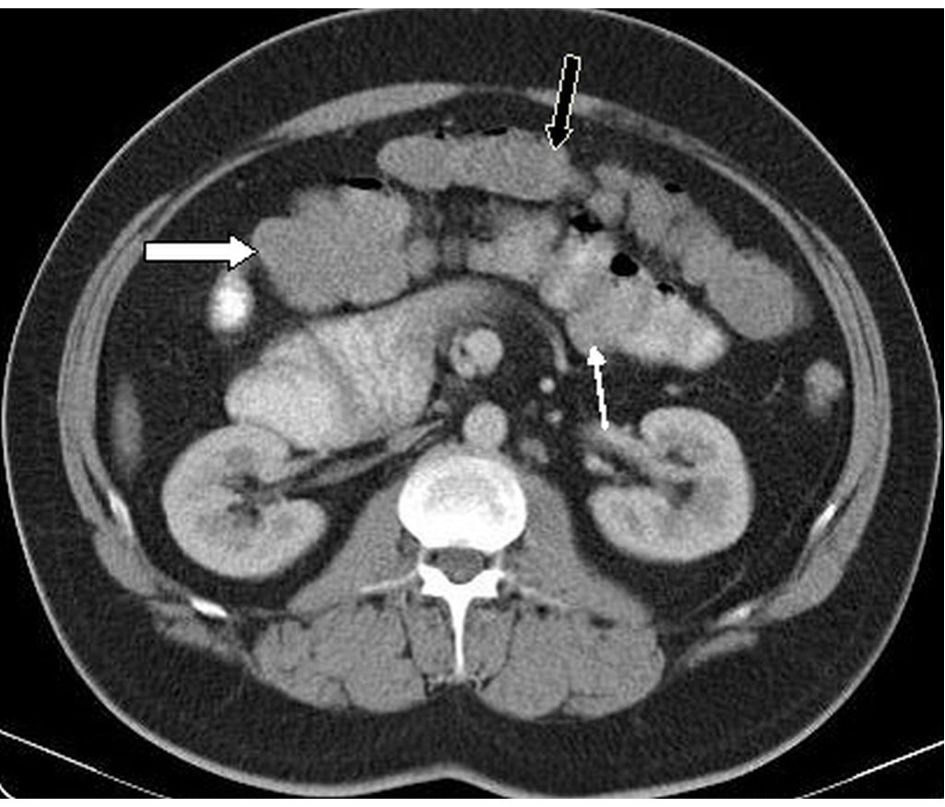
Figure 1. In axial CT imaging at infraumbilical level cecum and appendix was seen in the left lower abdominal quadrant (white arrow). Collabed dessendan colon was seen in lateral of cecum (black arrow).
| Journal of Medical Cases, ISSN 1923-4155 print, 1923-4163 online, Open Access |
| Article copyright, the authors; Journal compilation copyright, J Med Cases and Elmer Press Inc |
| Journal website http://www.journalmc.org |
Case Report
Volume 5, Number 5, May 2014, pages 267-269
Recurrent Vomiting and Epigastric Pain Attacks in an Adult Patient With Colonic Malrotation
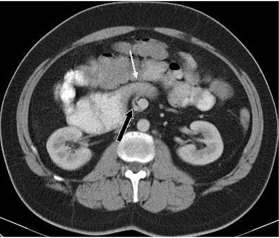
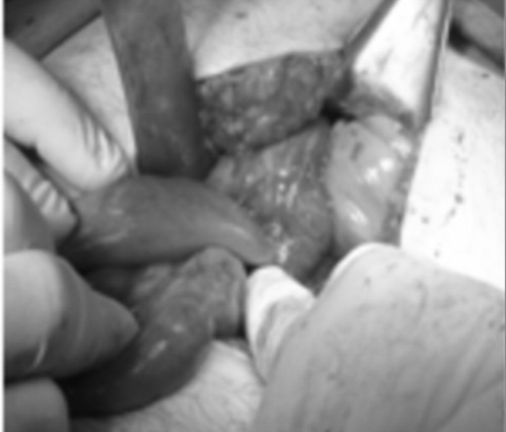
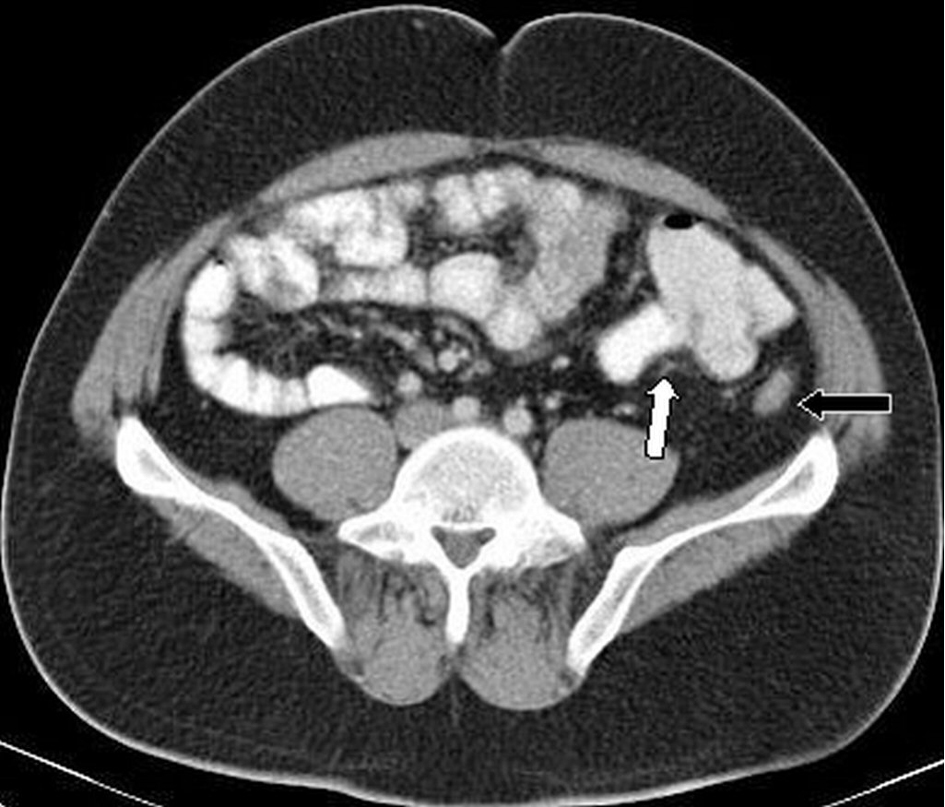
Figures