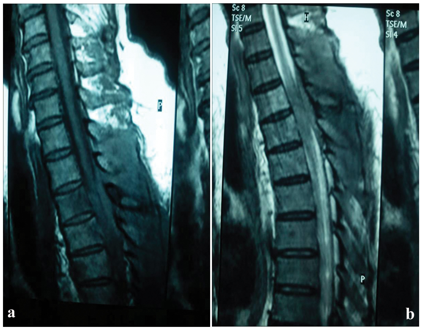
Figure 1. Sagittal post-contrast T1-weighted image of the thoracic spine reveals a lesion in the posterior (increased signal intensity) epidural region compressing the cord (a). Sagittal T2-weighted image of the thoracic spine at the same level depicts the same lesion in the posterior epidural region with low signal intensity (b).
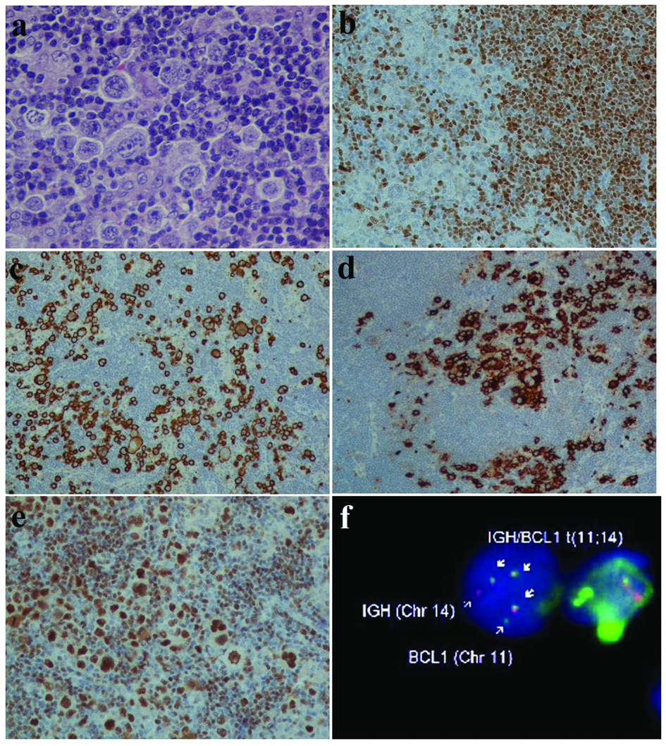
Figure 2. Neoplastic mantle cells and RS-like cells (a). Mantle cells positive to cyclinD1 (b) and RS cells immunoreactive to CD30 (c) and CD15 (d). Oct2 strong positivity of MCL and HRS cells (e). FISH method with IGH (red) and BCL1 (or CCND1) (green) gene probes revealing IGH/CCND1 gene fusion (arrows) (f). (a: HE, × 400; b, e: IHC, × 200; c, d: IHC, × 100).