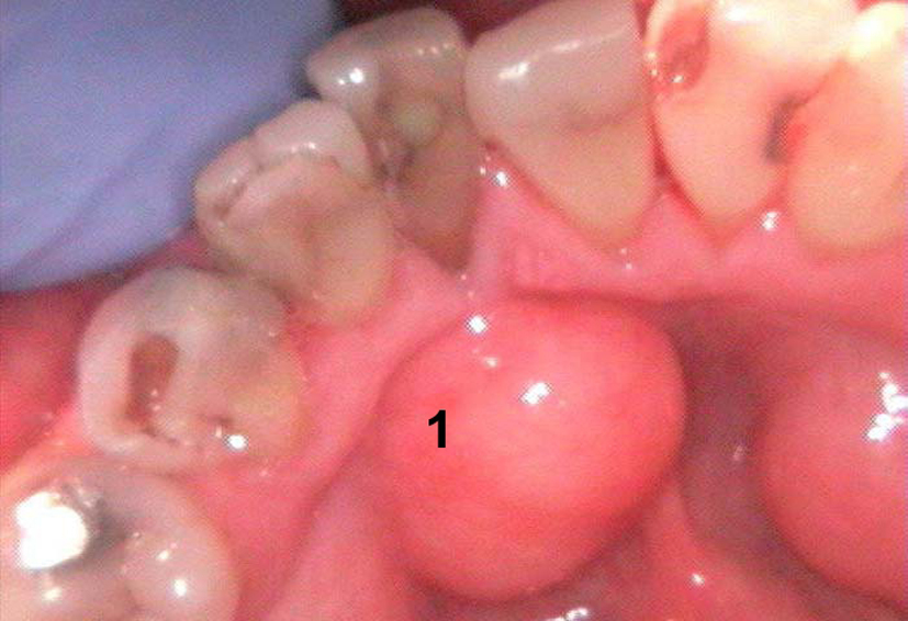
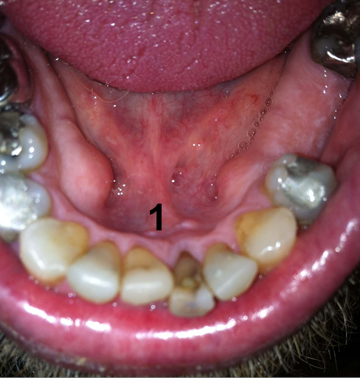
Figure 1. Intraoral photograph showing a well-defined mass (1) in the anterior floor of the mouth.
| Journal of Medical Cases, ISSN 1923-4155 print, 1923-4163 online, Open Access |
| Article copyright, the authors; Journal compilation copyright, J Med Cases and Elmer Press Inc |
| Journal website http://www.journalmc.org |
Case Report
Volume 5, Number 6, June 2014, pages 341-343
Chondrolipoma Arising in the Floor of the Mouth and Review of the Literature: A Case Report
Figures
Table
| Author | Location | Gender | Age in years |
|---|---|---|---|
| Pitman and Bell [2] | Masseter | Female | 51 |
| Berg and Gorsky [3] | Tongue | Male | 69 |
| Bezerra et al [4] | Tongue | Female | 68 |
| Goel et al [5] | Tongue | Female | 36 |
| Hietnen and Makinen [6] | Tongue | Female | 68 |
| Maes and Eulderink [7] | Tongue | Male | 47 |
| Nonaka et al [8] | Tongue | Male | 30 |
| Shabbir and Greenwood [9] | Tongue | Male | 71 |
| Allard et al [10] | Lower lip | Female | 69 |
| McAndrew and Greenspan [11] | Lower lip | Male | 72 |
| Fujimura and Enomoto [12] | Tongue | Male | 56 |
| Szudrowicz and Jakobi-Roz [13] | Lower lip | Male | 52 |
| Batchvarova et al [14] | Tongue | Male | 14 |
| Current case | Floor of mouth | Male | 56 |