Journal of Medical Cases, ISSN 1923-4155 print, 1923-4163 online, Open Access
Article copyright, the authors; Journal compilation copyright, J Med Cases and Elmer Press Inc
Journal website http://www.journalmc.org

Case Report

Volume 5, Number 4, April 2014, pages 253-255


Pigmented Villonodular Synovitis Masquerading as Metastasis on PET Imaging

Figures

Figure 1.
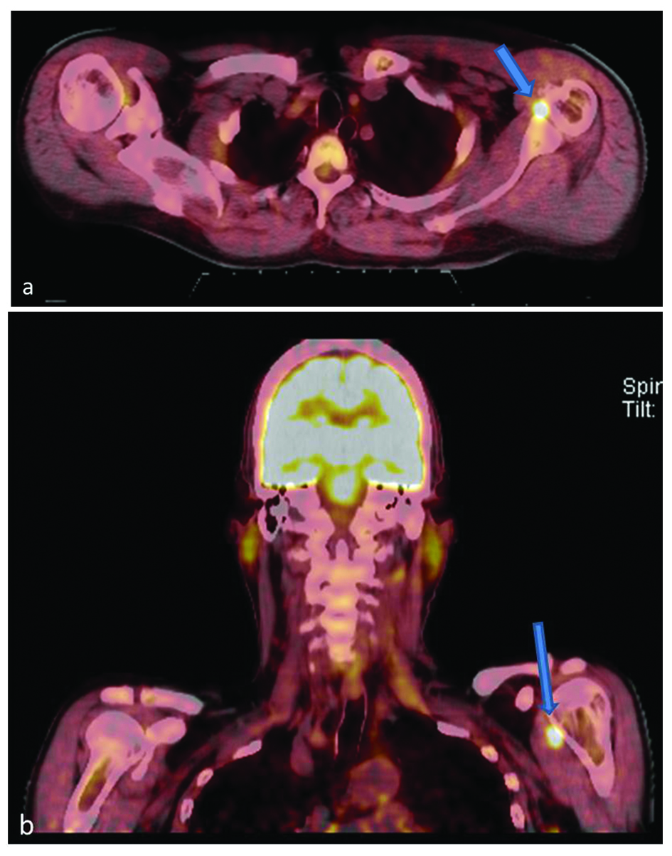
Figure 1. 18F-FDG PET axial (a) and coronal (b) images show a focal region of increased uptake (arrows) just medial to the left humeral head. The SUV maximum of this lesion was 11.6.
Figure 2.
Figure 2. Axial STIR (a) and T1 (b) magnetic resonance images show a focal region of intermediate STIR and low T1 signal (arrows) medial to the humerus corresponding to the focus of increased uptake on PET scanning.
Figure 3.
Figure 3. Axial CT image during percutaneous biopsy showing the needle tip directed toward the area of interest.
Figure 4.
Figure 4. (a) Diff-Quick stained touch preparation shows multinucleated giant cells (short arrow) and macrophages (long arrow) within a background of abundant, small synovial-like cells. (b) The hematoxylin and eosin stained core biopsy tissue contains similar findings, in addition to hemosiderin (arrow).