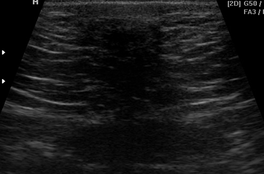
Figure 1. In B-mode ultrasonography (USG) a hypoechoic mass was observed, approximately 3 × 2 cm in size, with light acoustic shadow, irregular contours, and located vertically due to shrinking in the surrounding tissues. When the lesion was evaluated only with the USG findings, it had the properties of BI-RADS V appearance.
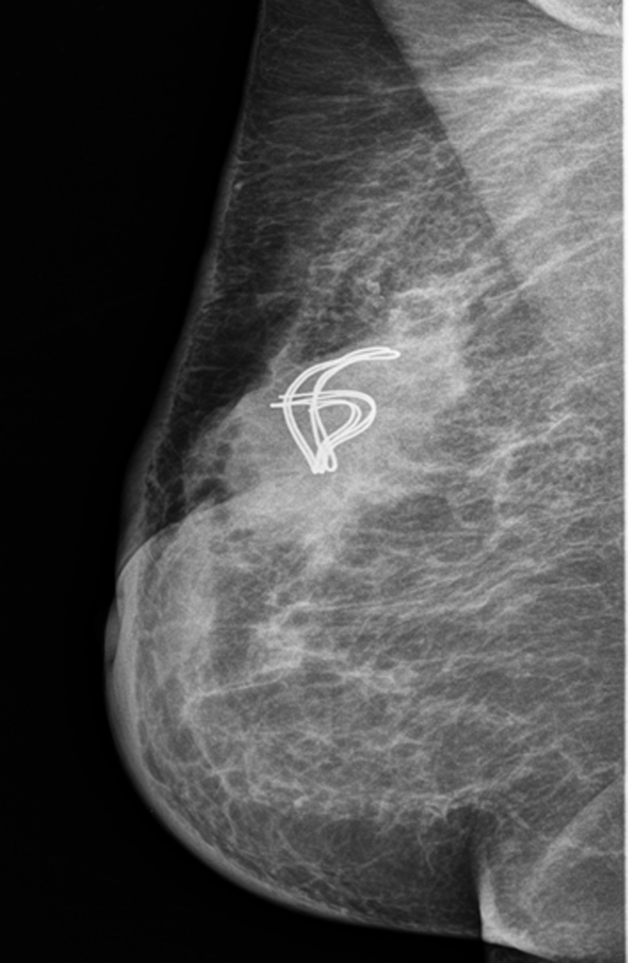
Figure 2. Meckel’s diverticulum after opening of the hernia sac.On the MLO radiograph, asymmetric density of equivalent intensity to breast glandular tissue in the surgical localisation of the right breast upper quadrant was observed. Within this area there was a twisted, linear appearance, which was thought to be suture material.
