Journal of Medical Cases, ISSN 1923-4155 print, 1923-4163 online, Open Access
Article copyright, the authors; Journal compilation copyright, J Med Cases and Elmer Press Inc
Journal website http://www.journalmc.org

Case Report

Volume 5, Number 3, March 2014, pages 163-166


An Incidentally Found True Portal Vein Aneurysm Secondary to Left Liver Lobe Aplasia in a Non-Cirrhotic Pregnant Woman Presented With Ovarian Torsion

Figures

Figure 1.
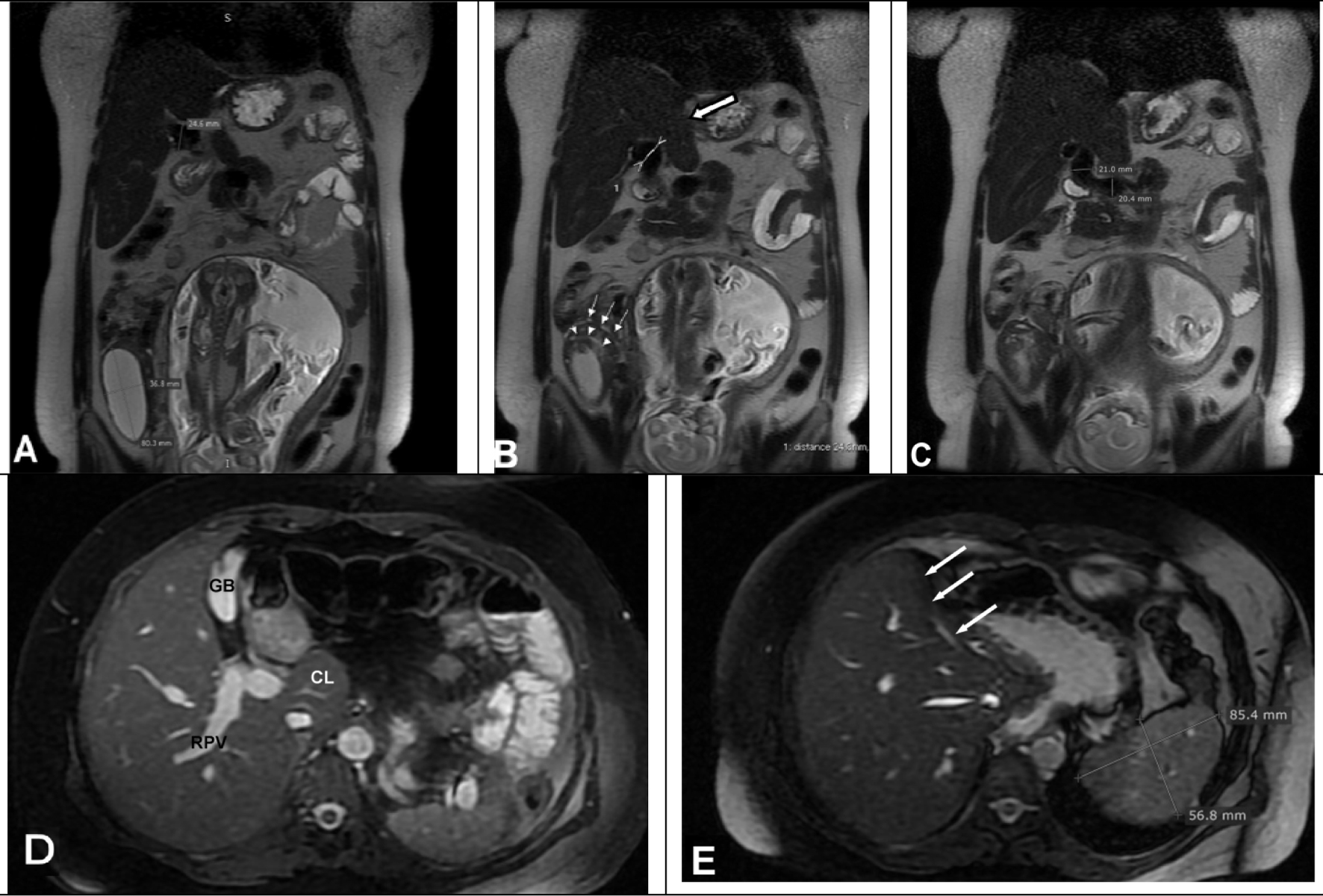
Figure 1. Coronal T2-weighted images (A, B) show a smooth edged hyperintense cystic lesion on right ovary and twisted salpinx (arrow heads and small arrows) was seen on superior aspect of the cyst. The left liver lobe was aplasic (big white arrow). Portal vein was dilated and diameters were measured 24 mm on distal portion, (C) 21 mm in the middle part and 20 mm in proximal portion. (D) Axial fat sat T2-weighted gradient echo image from mid level of liver shows right portal vein (RPV), gall bladder (GB) and caudat lobe (CL). (E) Axial gradient echo fat sat image from upper level of liver shows left liver lob was aplasic (arrows) and spleen sizes were in normal ranges.
Figure 2.
Figure 2. (A) Four-time anti-clockwise twisted ovary and tuba. (B) Cystic mass of 7 × 7 cm in size with hemorrhagic infarct. (C) Histopathologic view of ovarian tissues with hemorrhagic and (D) tuba with hemorrhagic infarct.
Figure 3.
Figure 3. (A) One month after the labor CDS images show hepatopedal blood flow and portal vein diameter was measured 21 mm. (B) Axial T1-weighted gradient echo and (C) gradient echo fat sat MR images show decreased portal vein diameters.