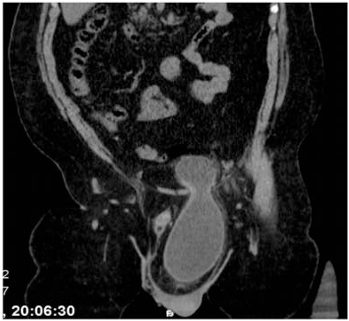
Figure 1. Coronal CT scan of left inguinal region, extending to the mesenteric adipose tissue reveals the bladder with left inguinal canal. Thickening of the bladder wall hernia was noticed at the level of the neck.
| Journal of Medical Cases, ISSN 1923-4155 print, 1923-4163 online, Open Access |
| Article copyright, the authors; Journal compilation copyright, J Med Cases and Elmer Press Inc |
| Journal website http://www.journalmc.org |
Case Report
Volume 5, Number 3, March 2014, pages 174-176
Clinical and Radiographic Findings of a Sliding Inguinoscrotal Bladder Hernia
Figures