Journal of Medical Cases, ISSN 1923-4155 print, 1923-4163 online, Open Access
Article copyright, the authors; Journal compilation copyright, J Med Cases and Elmer Press Inc
Journal website http://www.journalmc.org

Case Report

Volume 8, Number 2, February 2017, pages 63-66


Acute Retropharyngeal Calcific Tendinitis or Calcium Hydroxyapatite Deposits in Longus Colli Muscle

Figures

Figure 1.
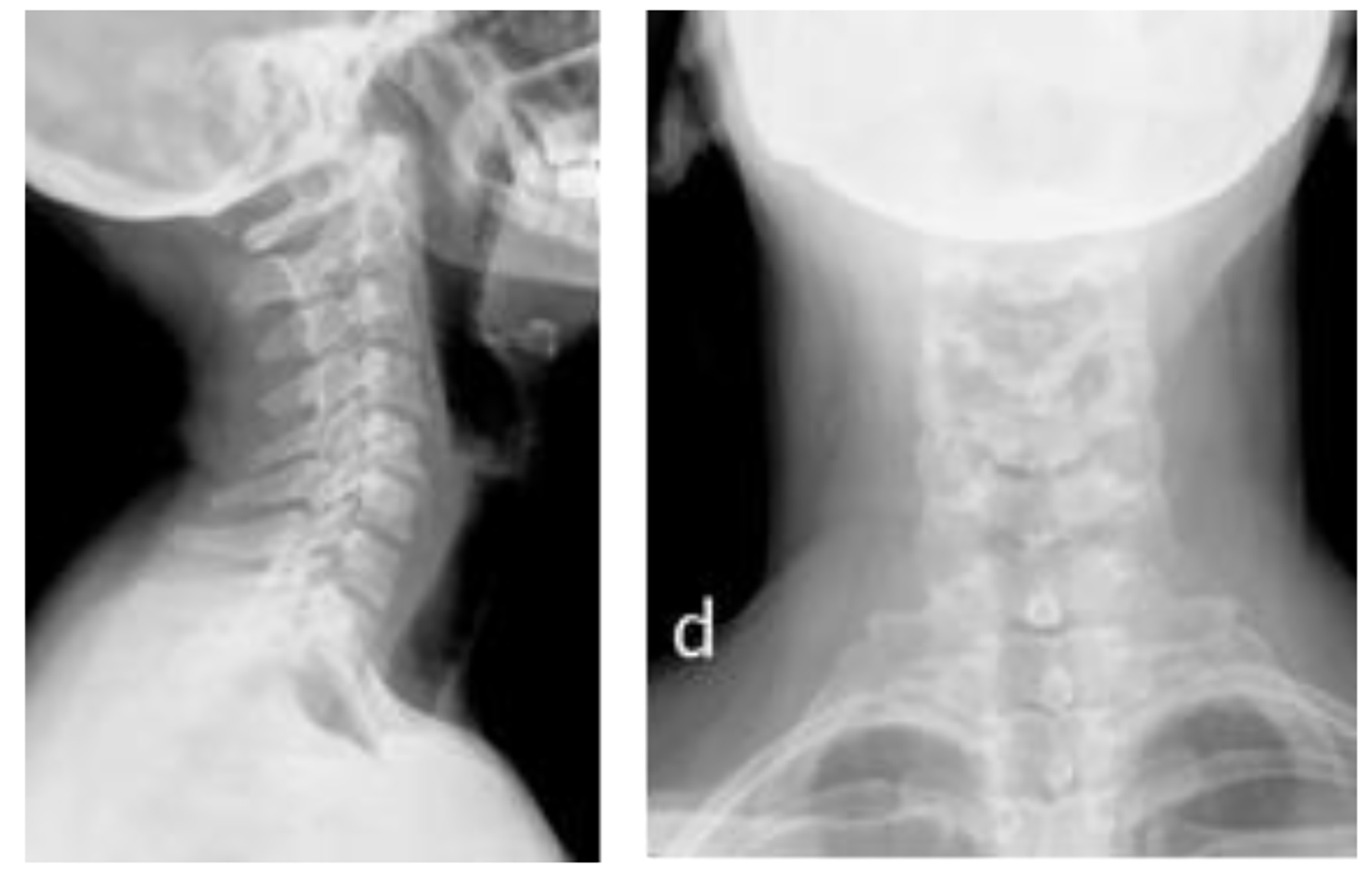
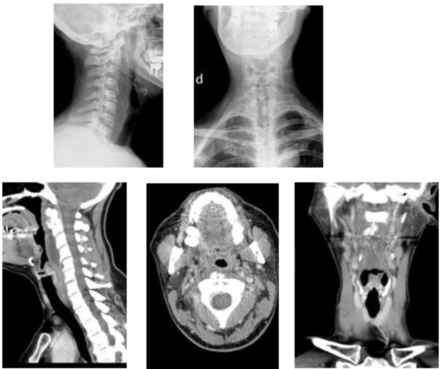
Figure 1. Two images of cervical plain on top. The first side showing rectification of physiological lordosis next to the prevertebral calcifications at level C1-C2 and edematization of the prevertebral space. Pictures of CT on the bottom showing moderate increase of prevertebral soft tissues at the height of C2, C3 and C4 and a tenuous calcification of irregular morphology, approximately 15 × 14.5 × 9.5 mm in intimate relation with the proximal portion of the longus colli muscle belly.
Figure 2.
Figure 2. Two simple cervical X-ray images are shown at 2 weeks of evolution. The first side shows again physiological lordosis and a disappearance of the prevertebral calcifications at level C1-C2 and decrease of edema of the prevertebral space.