Journal of Medical Cases, ISSN 1923-4155 print, 1923-4163 online, Open Access
Article copyright, the authors; Journal compilation copyright, J Med Cases and Elmer Press Inc
Journal website http://www.journalmc.org

Case Report

Volume 5, Number 1, January 2014, pages 45-48


A Case of an Tubular Foreign Body in the Paranasal Sinus After Penetrating Orbital Injury

Figures

Figure 1.
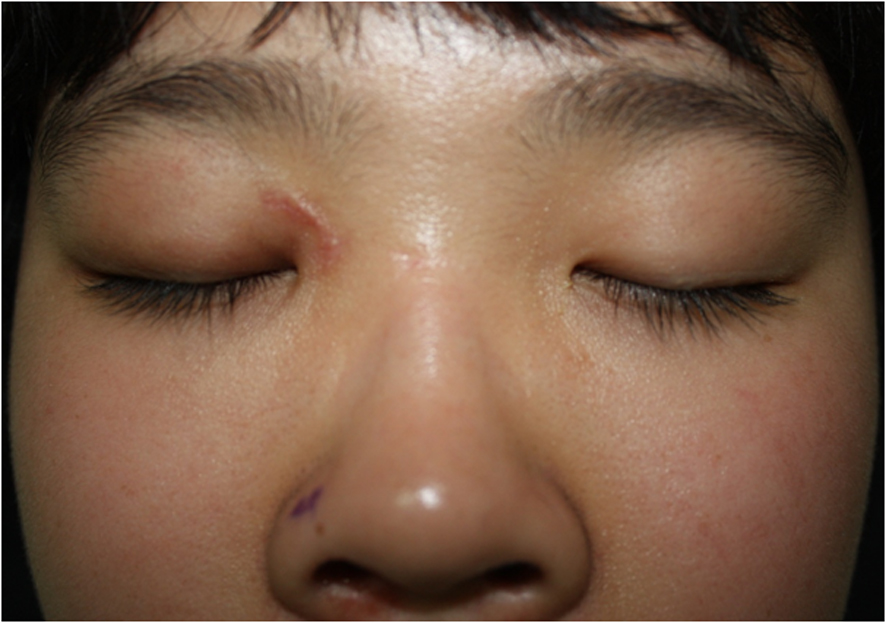
Figure 1. Right medical canthus scar tissue (about a 2 cm sized laceration).
Figure 2.
Figure 2. Paranasal sinus CT axial view. There is an air-filled non-enhancing tubular structure extending from the right orbit to the posterior wall of the sphenoid sinus.
Figure 3.
Figure 3. Paranasal sinus CT (sagittal view). There is an air-filled non-enhancing tubular structure which is located in the right orbit and extends to the posterior wall of the sphenoid sinus.
Figure 4.
Figure 4. Removed foreign body (8 x 1 cm sized ball pen).
Figure 5.
Figure 5. Postoperative paranasal CT findings (axial and coronal view).