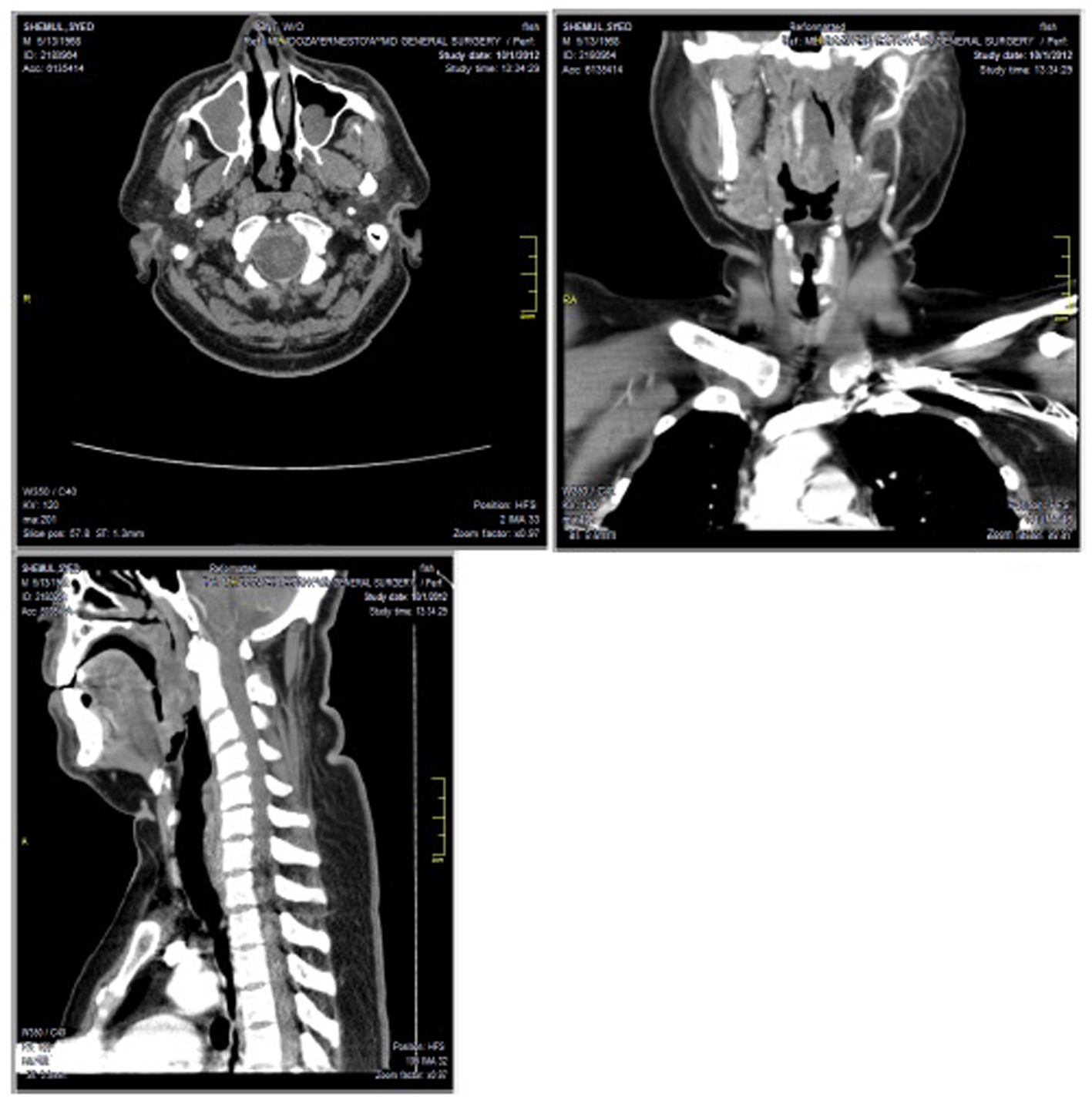
Figure 1. CT scan of transverse, coronal and saggital views demonstrating mass in nasopharynx.
| Journal of Medical Cases, ISSN 1923-4155 print, 1923-4163 online, Open Access |
| Article copyright, the authors; Journal compilation copyright, J Med Cases and Elmer Press Inc |
| Journal website http://www.journalmc.org |
Case Report
Volume 5, Number 2, February 2014, pages 58-61
Recurrent Rhinosporidiosis
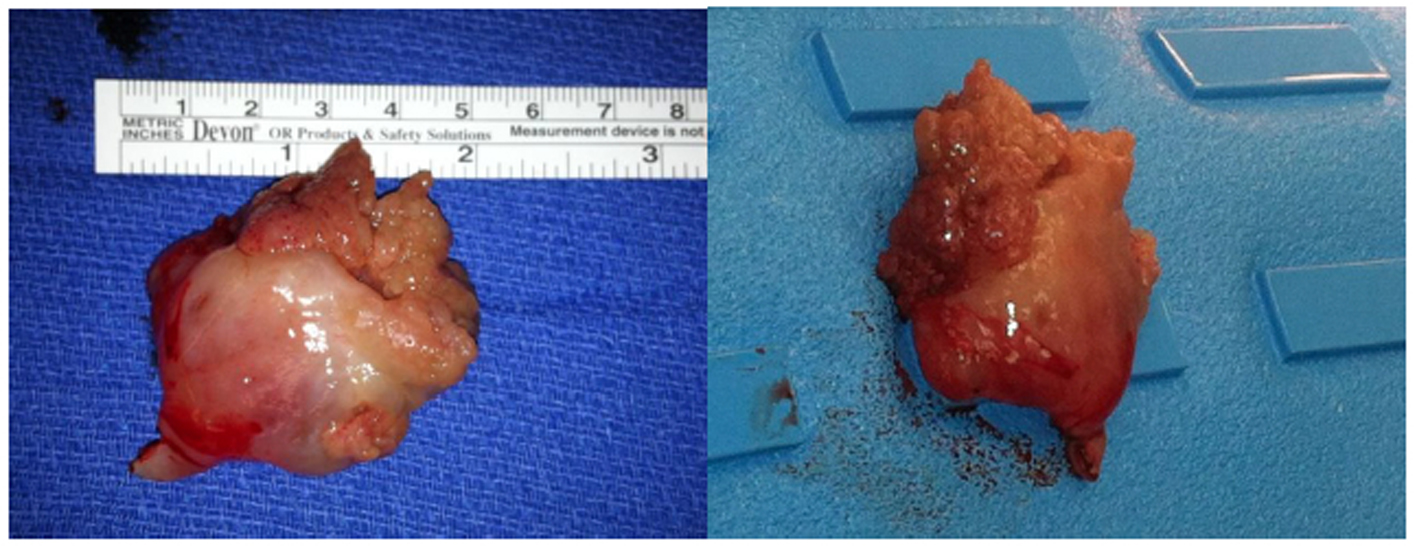
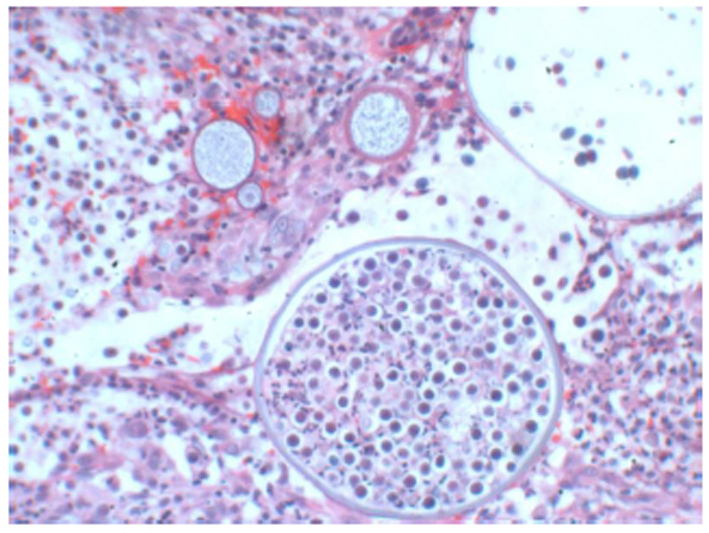
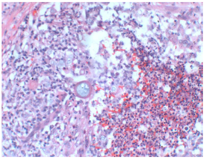
Figures