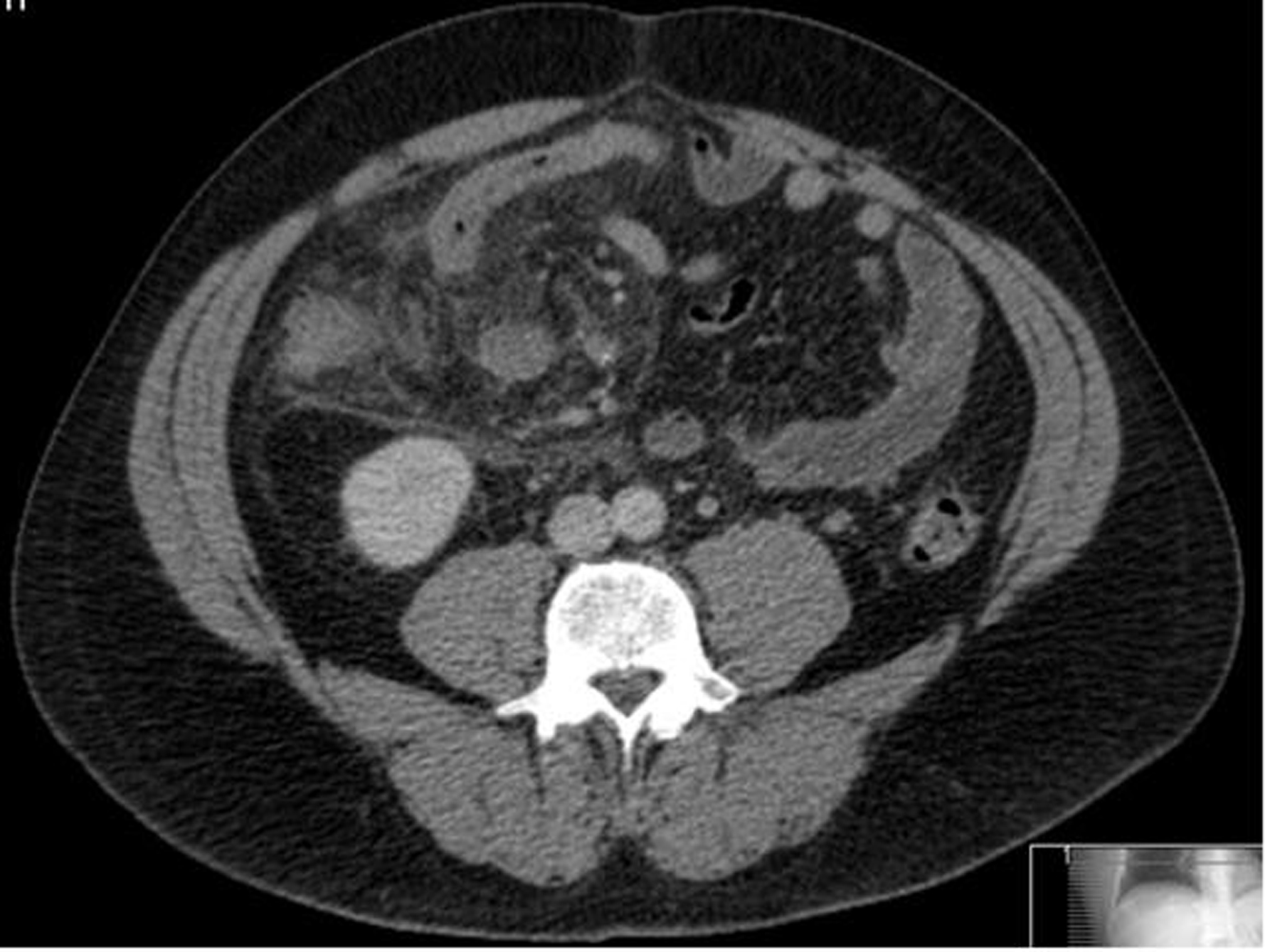
Figure 1. This is an axial section from the CT scan demonstrating the large amount of inflammatory change within the right iliac fossa.
| Journal of Medical Cases, ISSN 1923-4155 print, 1923-4163 online, Open Access |
| Article copyright, the authors; Journal compilation copyright, J Med Cases and Elmer Press Inc |
| Journal website http://www.journalmc.org |
Case Report
Volume 2, Number 6, December 2011, pages 296-299
Fish Bone Perforation Mimicking Acute Appendicitis
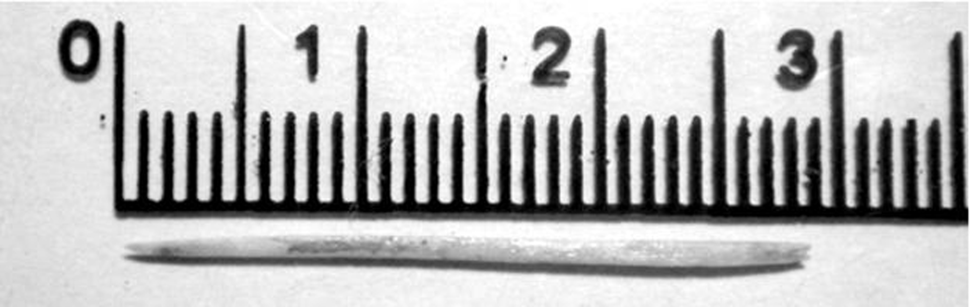
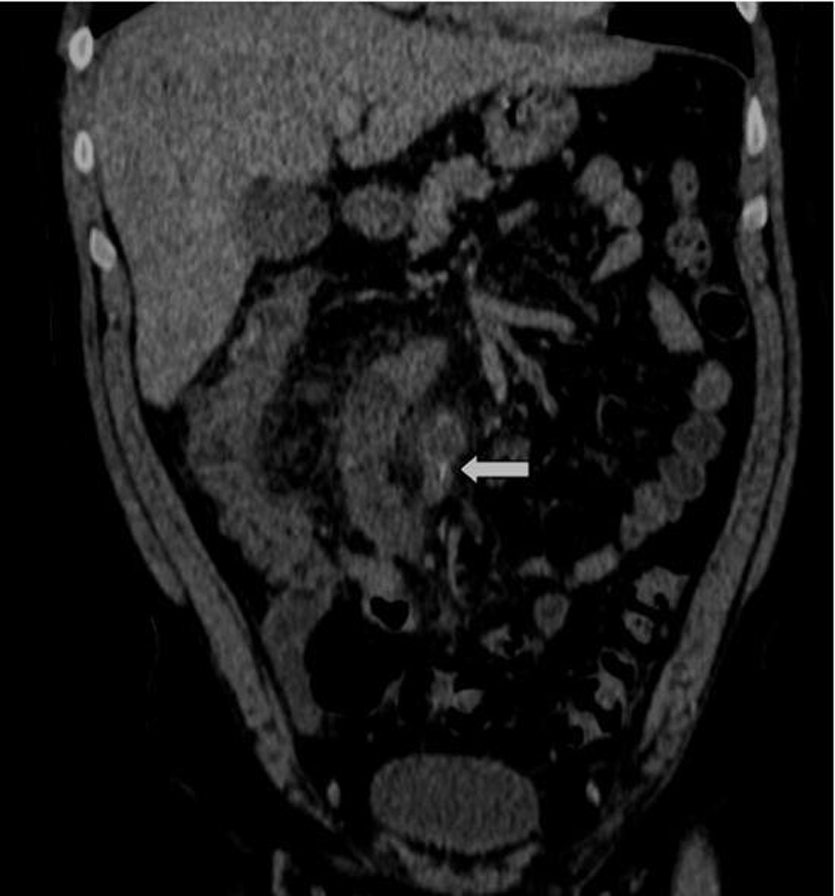
Figures