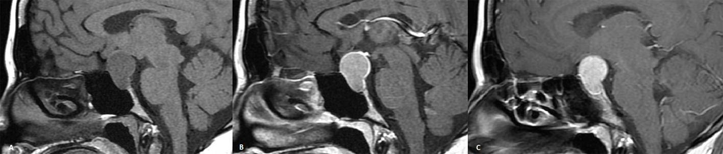
Figure 1. A) Sagittal pre-contrast T1WI shows mild hypointensity, B) post-contrast T1WI MRI showing peripheral contrast enhancement in the first series, and C) homogen contrast enhancement is observed in the subsequent series.
| Journal of Medical Cases, ISSN 1923-4155 print, 1923-4163 online, Open Access |
| Article copyright, the authors; Journal compilation copyright, J Med Cases and Elmer Press Inc |
| Journal website http://www.journalmc.org |
Case Report
Volume 4, Number 11, November 2013, pages 719-721
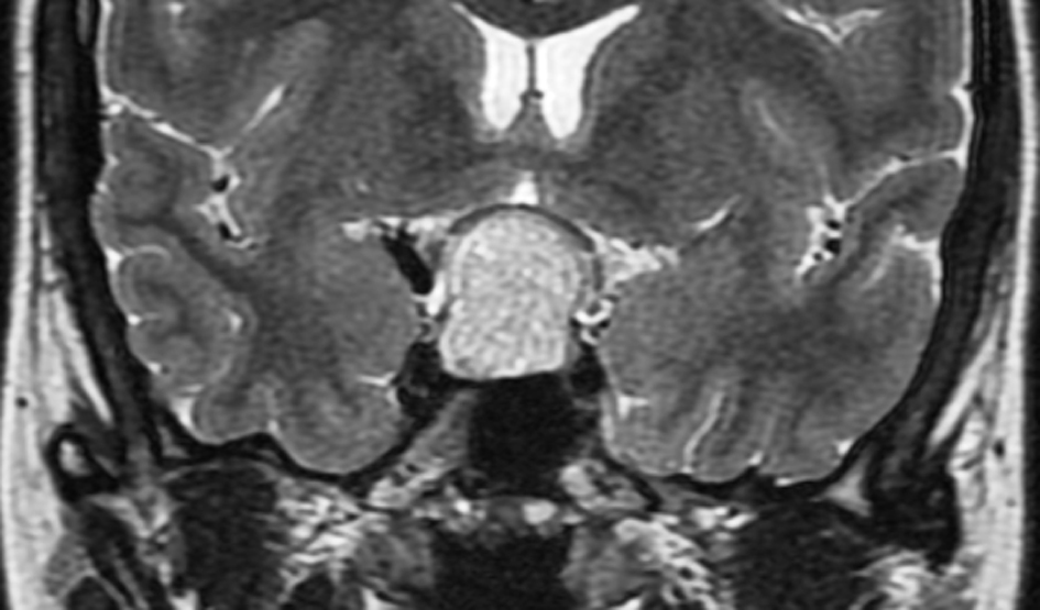
Intrasellar Cavernous Hemangioma: MRI Findings of a Very Rare Lesion
Figures