Journal of Medical Cases, ISSN 1923-4155 print, 1923-4163 online, Open Access
Article copyright, the authors; Journal compilation copyright, J Med Cases and Elmer Press Inc
Journal website http://www.journalmc.org

Case Report

Volume 4, Number 10, October 2013, pages 679-681


A Cavitary Conundrum

Figures

Figure 1.
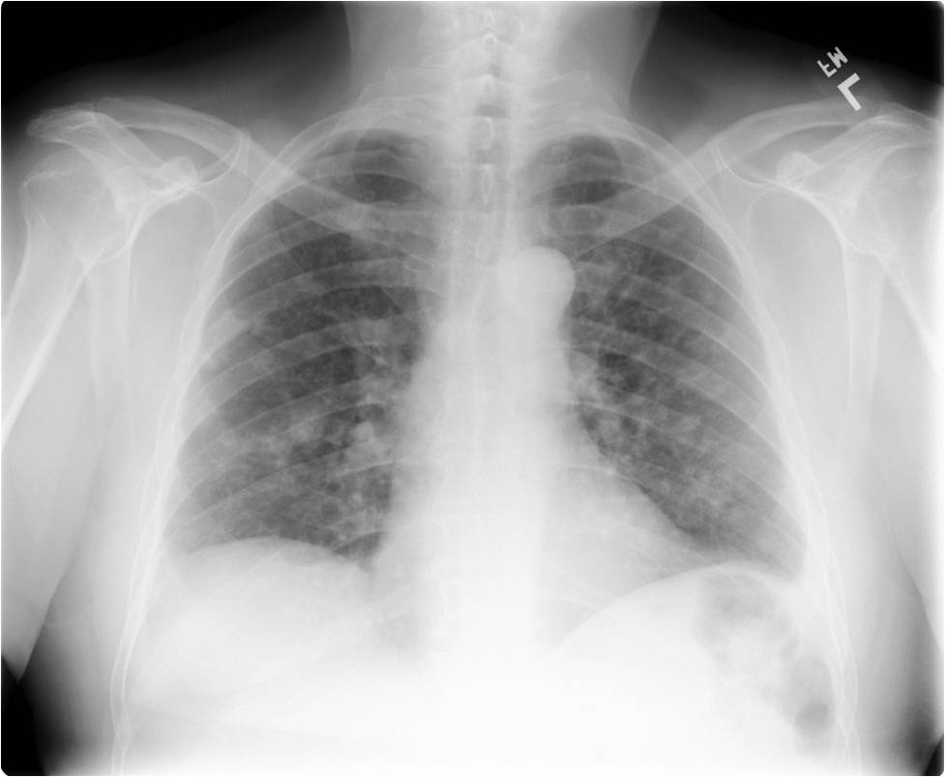
Figure 1. Frontal radiograph of the chest showing multiple bilateral pulmonary nodules.
Figure 2.
Figure 2. (a) Axial and (b) coronal CT images in lung windows show multiple bilateral pulmonary nodules of varying sizes, some cavitary.
Figure 3.
Figure 3. (a) Histology from core biopsy demonstrates malignant glands consistent with an adenocarcinoma (hemotoxylin-esosin stain 10 ×). (b) Staining for prostate acid phosphatase (PSAP) confirms the diagnosis of metastatic prostatic adenocarcinoma (immunostain for PSAP 20 ×).