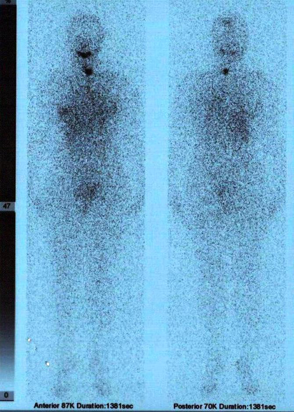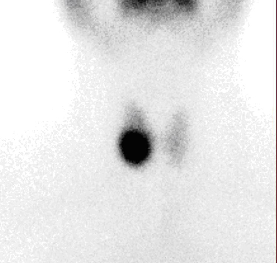
Figure 1. 99mTc scintigraphy showing a hot nodule at the right thyroid region.
| Journal of Medical Cases, ISSN 1923-4155 print, 1923-4163 online, Open Access |
| Article copyright, the authors; Journal compilation copyright, J Med Cases and Elmer Press Inc |
| Journal website http://www.journalmc.org |
Case Report
Volume 4, Number 10, October 2013, pages 686-688
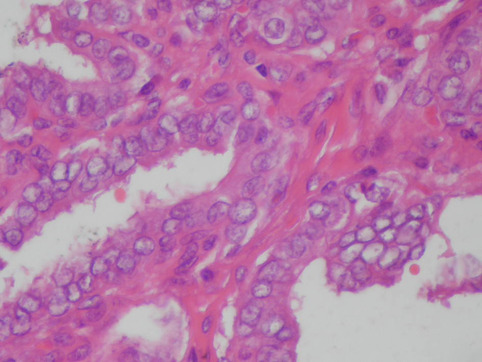
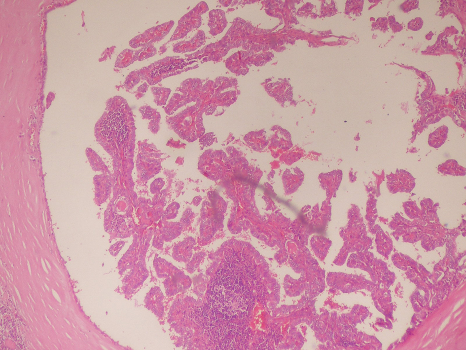
Papillary Thyroid Carcinoma Developments After Radioactive Iodine Treatment for Toxic Adenoma: A Case Report
Figures