Figures
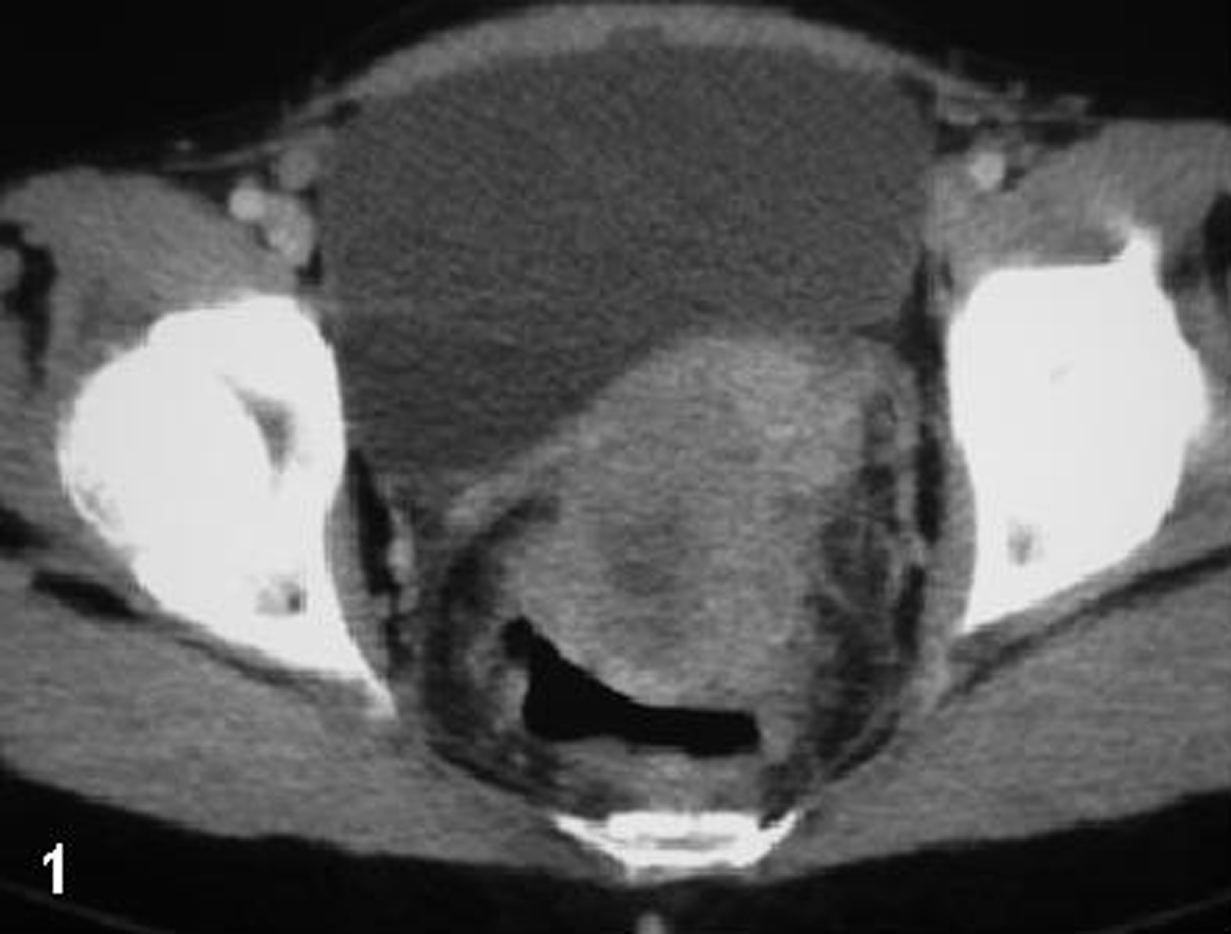
Figure 1. CT of the pelvis: a space occupying lesion originating from the rectal wall invading the uterine corps.
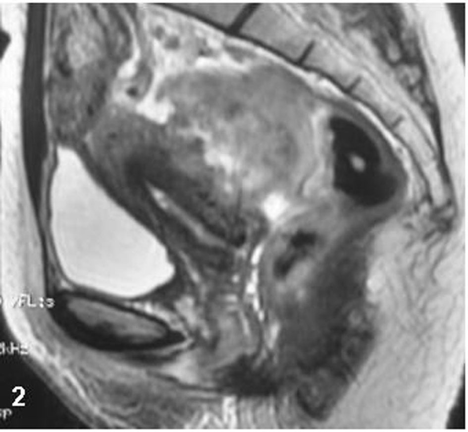
Figure 2. Sagittal MR imaging of the pelvis, T2W sequence: an inhomogeneous massive lesion originating from the anterior rectal wall invades the myometrium.
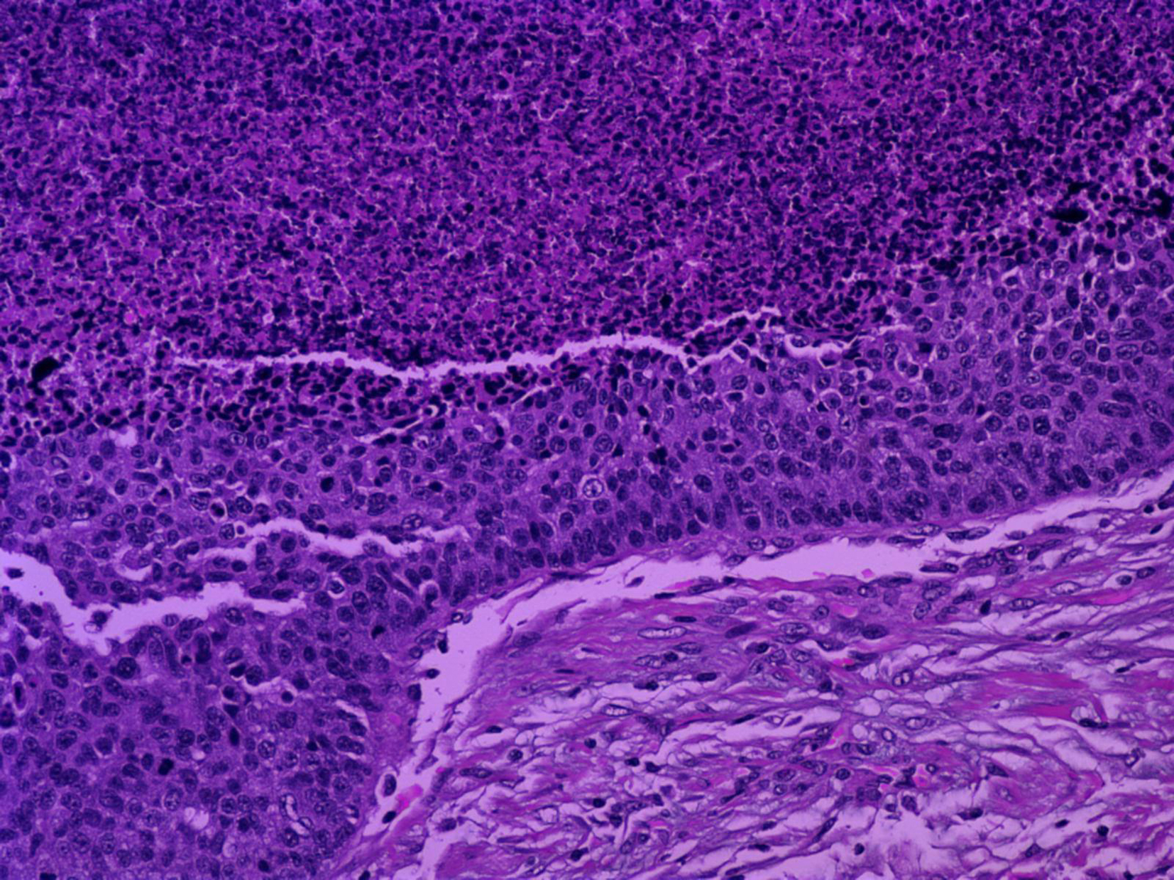
Figure 3. (Hematoxylin and Eosin/X20): Neoplasm in the better differentiated foci characterized by the formation of tumor nests with central necrosis and peripheral palisading of small to medium sized malignant cells.
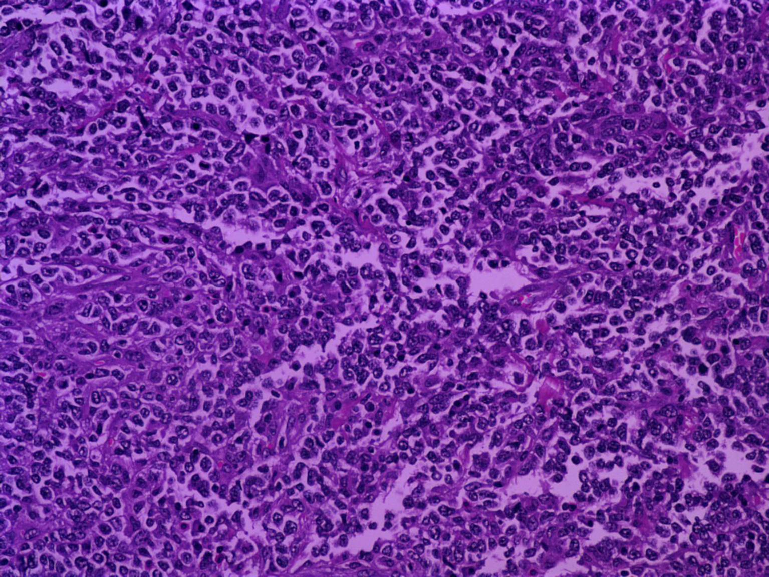
Figure 4. (Hematoxylin and Eosin/X20): Undifferentiated areas within tumor, characterized by diffusely distributed malignant cells.
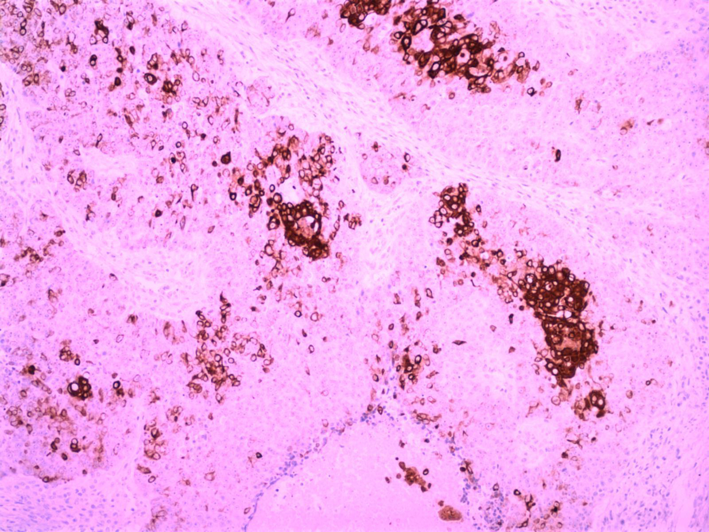
Figure 5. (immunolabeling for CK34BE12/X10): Immunoreactivity to CK34BE12 from the better differentiated neoplastic cells.
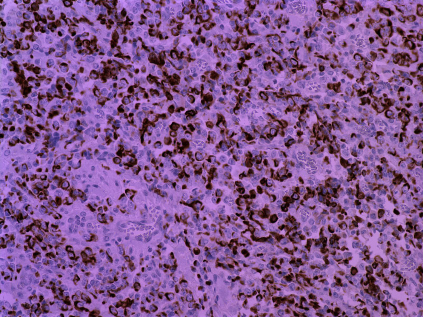
Figure 6. (immunolabeling for CK18/ X20): CK18 expression from undifferentiated malignant cells.
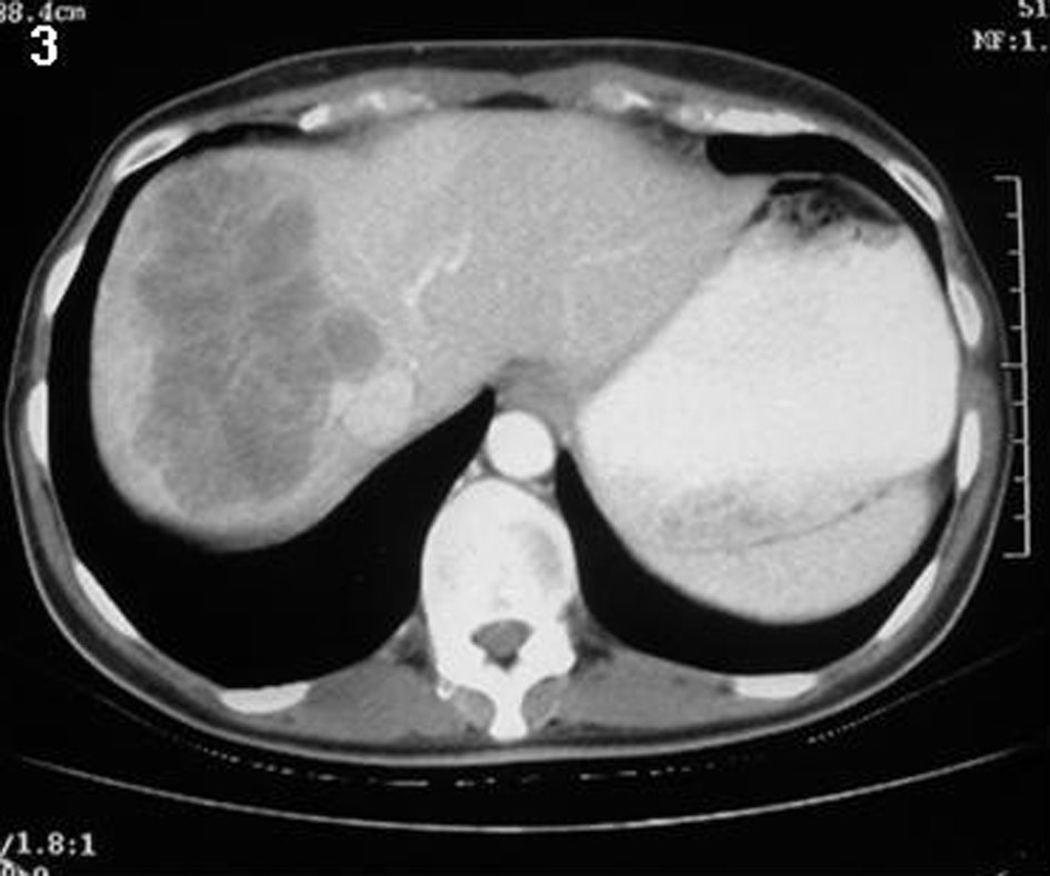
Figure 7. An abdominal CT revealed a large metastatic lesion of the right lobe of the liver.