Journal of Medical Cases, ISSN 1923-4155 print, 1923-4163 online, Open Access
Article copyright, the authors; Journal compilation copyright, J Med Cases and Elmer Press Inc
Journal website http://www.journalmc.org

Case Report

Volume 4, Number 9, September 2013, pages 616-619


Diagnosis of Median Arcuate Ligament Syndrome on Multidetector Computed Tomography

Figures

Figure 1.
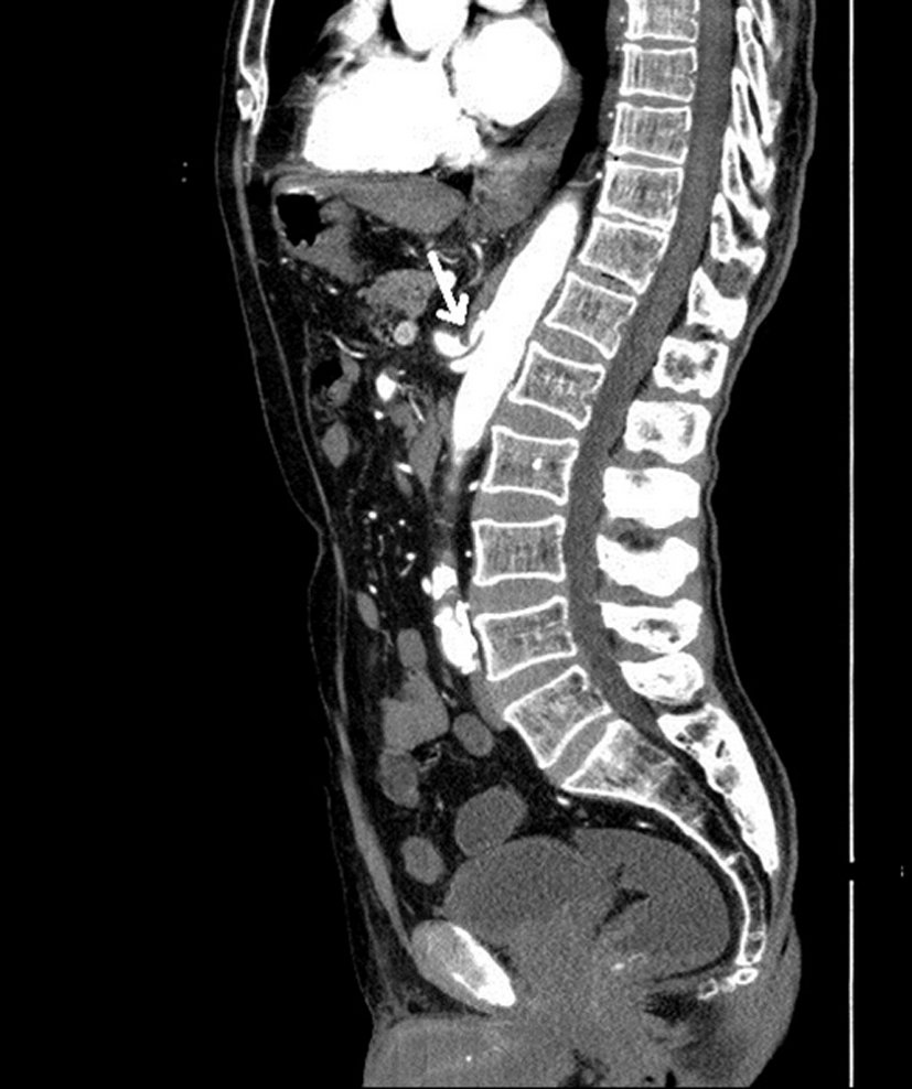
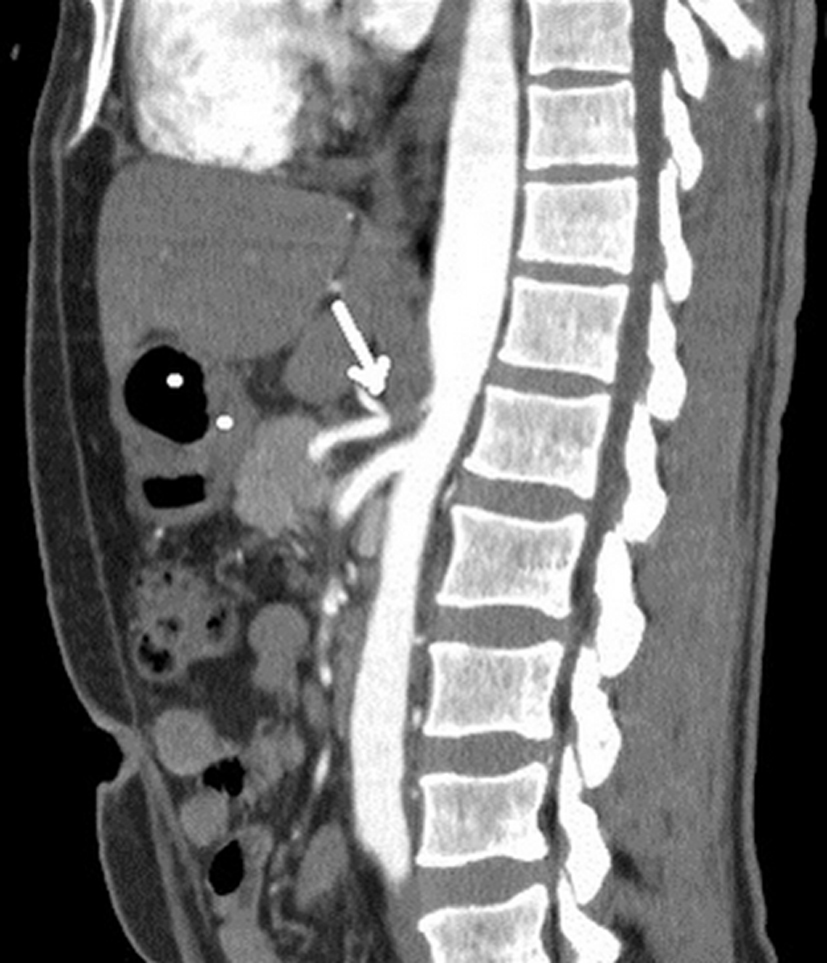
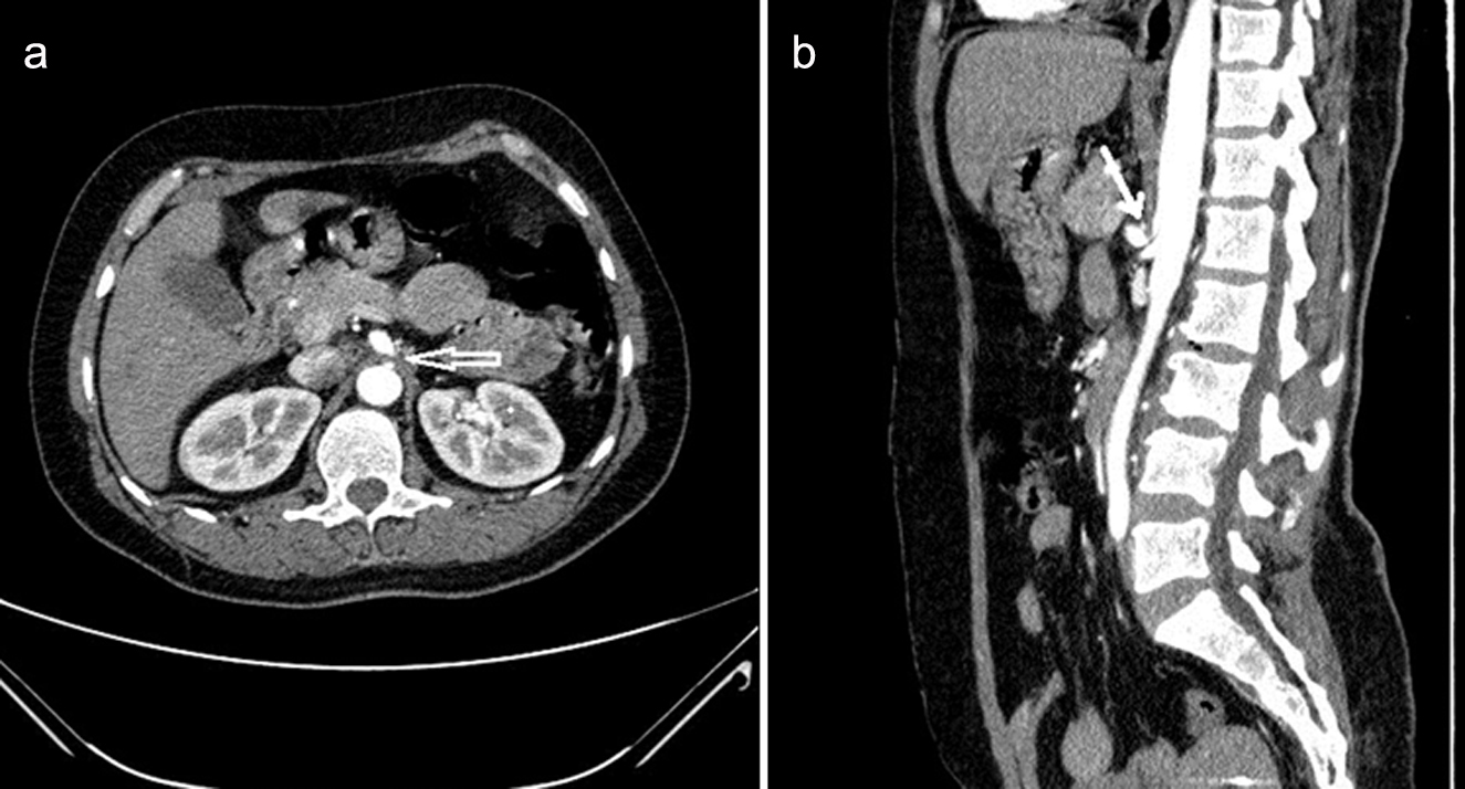
Figure 1. Reformatted axial (a) and sagittal (b) images (in the 39-year-old woman with MAL syndrome) show the compression of the celiac artery by the hypertrophic MAL, a narrowing in the celiac trunk lumen, and a poststenotic dilatation (arrow). The sagittal image shows a typical hooked appearance of the proximal celiac trunk.
Figure 2.
Figure 2. The reformatted sagittal CT image (in the 77-year-old man with MAL syndrome) shows the compression of the celiac artery by the hypertrophic MAL and a narrowing in the celiac trunk lumen (arrow). The sagittal image shows a typical hooked appearance of the proximal celiac artery.
Figure 3.
Figure 3. The reformatted sagittal CT image (in the 73-year-old woman with MAL syndrome) shows the compression of the celiac artery by the hypertrophic MAL and a narrowing in the celiac trunk lumen (arrow).