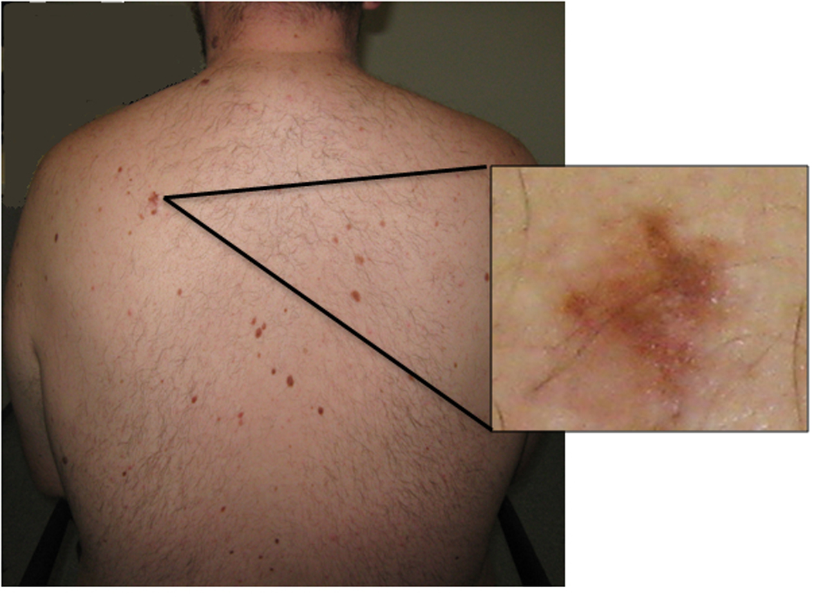
Figure 1. Photograph showing the back of the patient. Multiple pigmented dysmorphic nevi are evident. Inset: higher power view of a dysmorphic nevus.
| Journal of Medical Cases, ISSN 1923-4155 print, 1923-4163 online, Open Access |
| Article copyright, the authors; Journal compilation copyright, J Med Cases and Elmer Press Inc |
| Journal website http://www.journalmc.org |
Case Report
Volume 4, Number 8, August 2013, pages 526-529
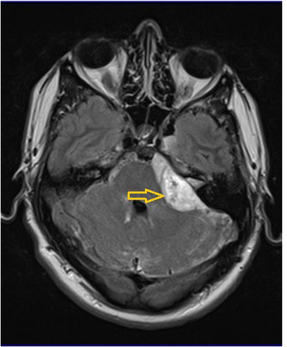
Primary Diffuse Leptomeningeal Gliomatosis Coincident With a Melanocytic Disorder
Figures