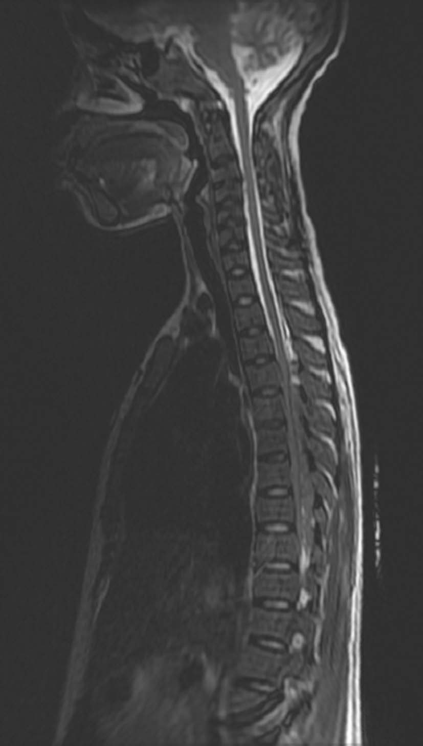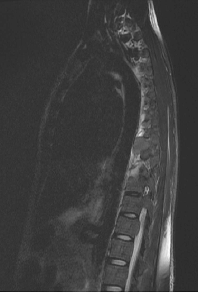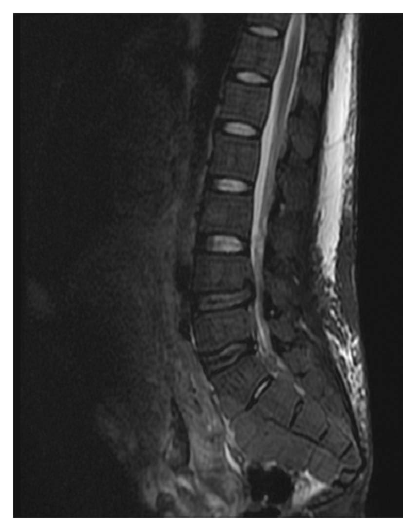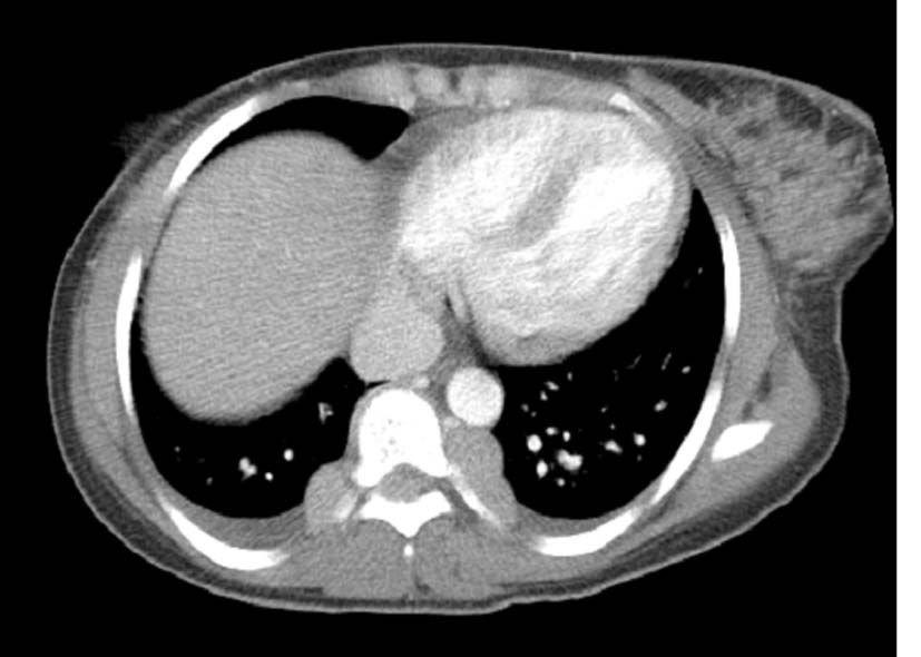
Figure 1. Bilateral para-spinal soft tissue masses consistent with extra-medullary hematopoietic tissue.
| Journal of Medical Cases, ISSN 1923-4155 print, 1923-4163 online, Open Access |
| Article copyright, the authors; Journal compilation copyright, J Med Cases and Elmer Press Inc |
| Journal website http://www.journalmc.org |
Case Report
Volume 2, Number 2, April 2011, pages 58-61
Beta-Thalassemia Presenting as an Acute Neurological Complication: A Case Report
Figures