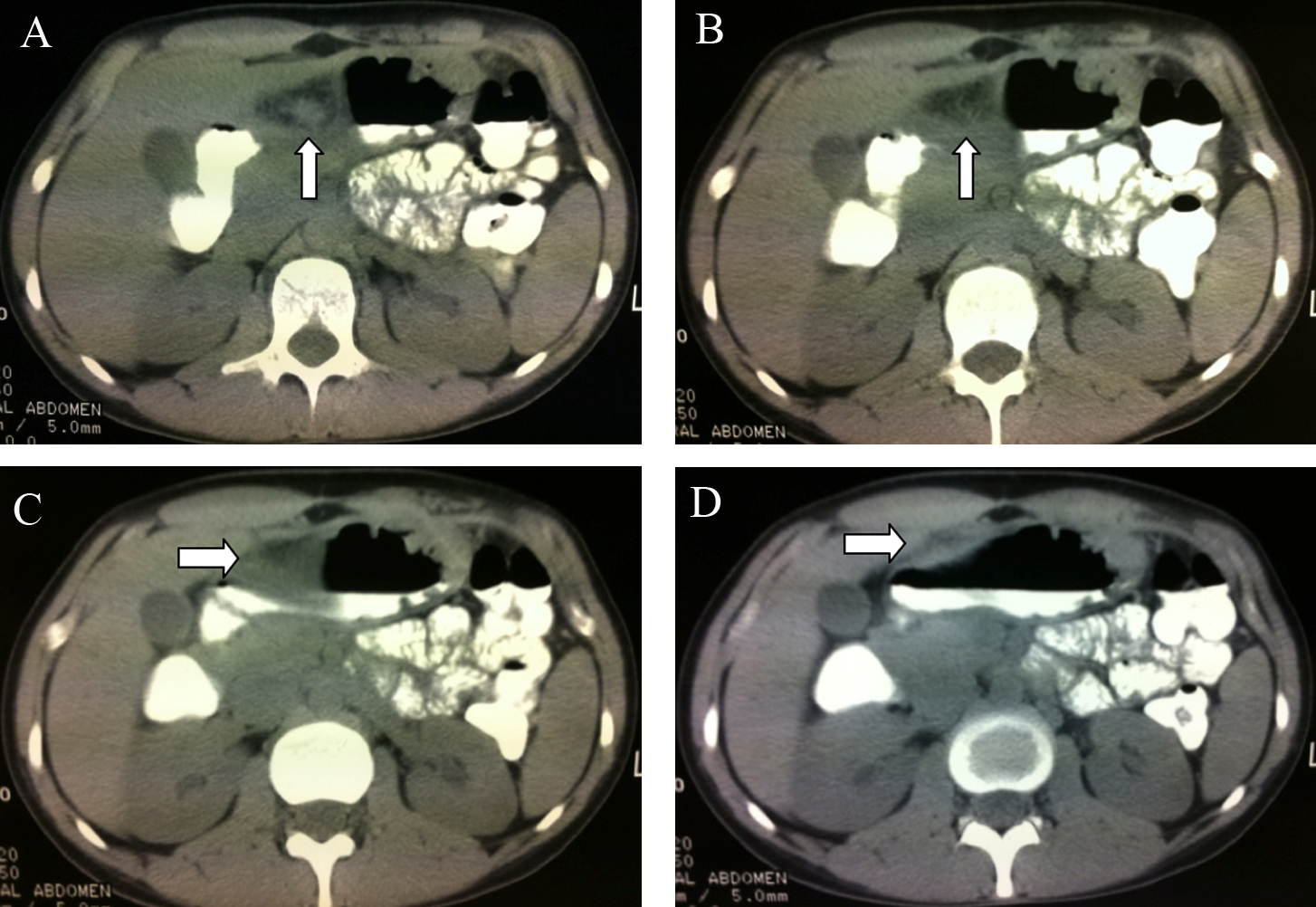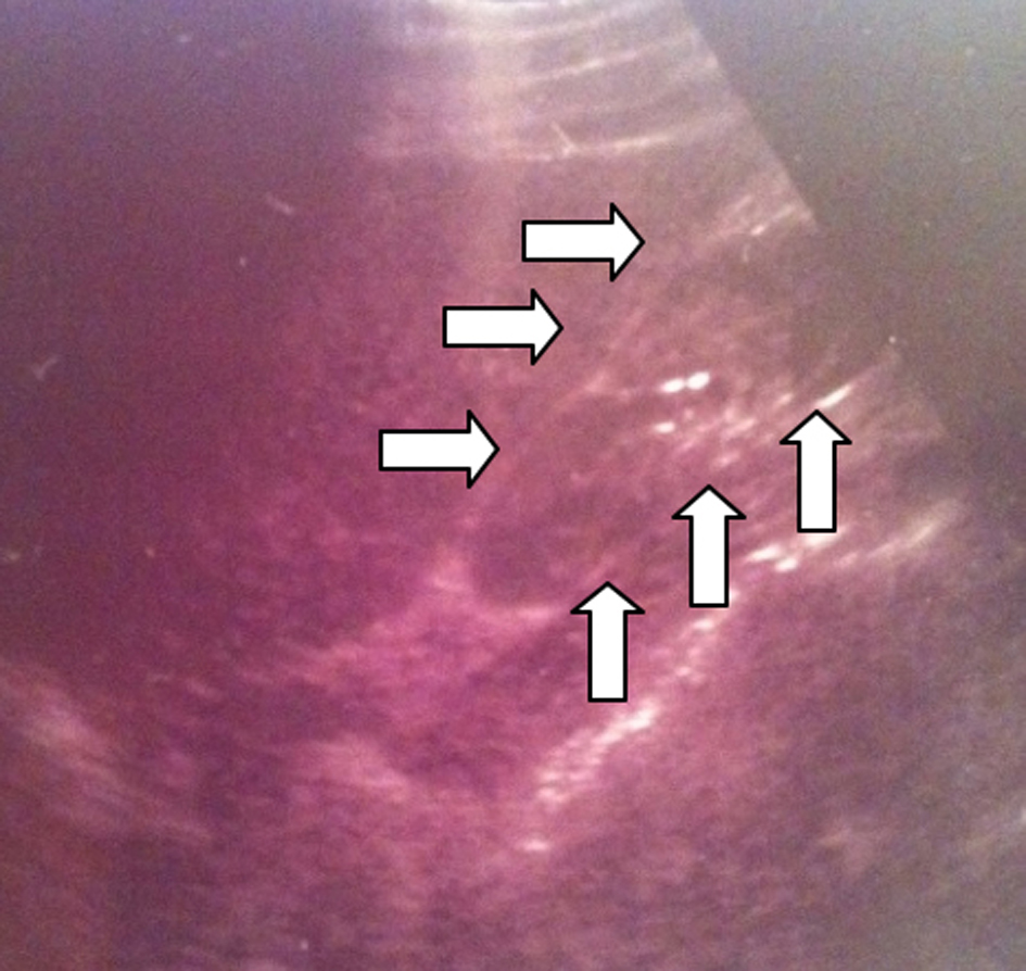
Figure 1. The abdominal U/S was evaluated as uncontributive, however a well circumscribed moderately hyperechoic formation in the anatomic location of the lesser omentum can be noticed (arrows).
| Journal of Medical Cases, ISSN 1923-4155 print, 1923-4163 online, Open Access |
| Article copyright, the authors; Journal compilation copyright, J Med Cases and Elmer Press Inc |
| Journal website http://www.journalmc.org |
Case Report
Volume 4, Number 7, July 2013, pages 499-503
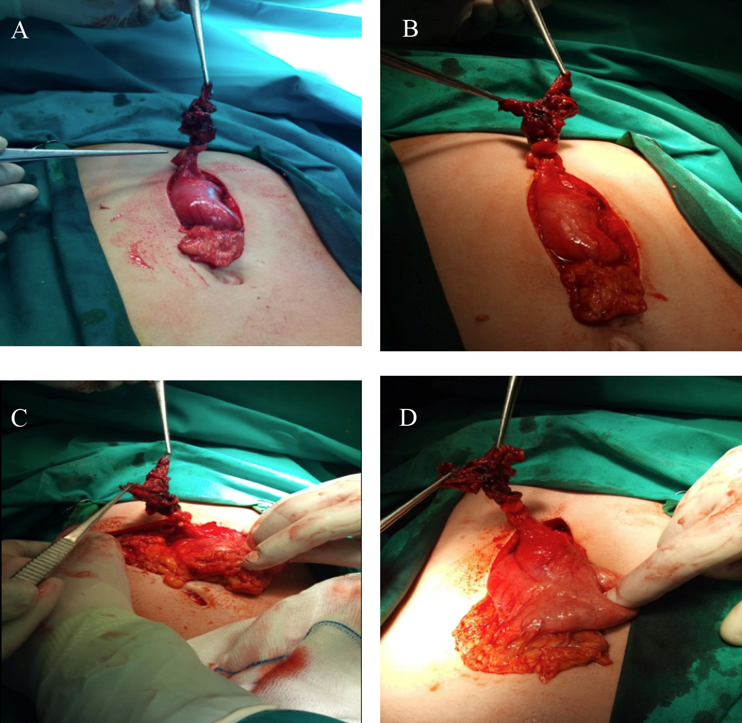
Primary Lesser Omentum Torsion - An Extremely Rare Cause of Acute Abdomen and a Very Uncommon Subtype of Intraperitoneal Focal Fat Infarction (IFFI): Case Report and Review of the Literature
Figures