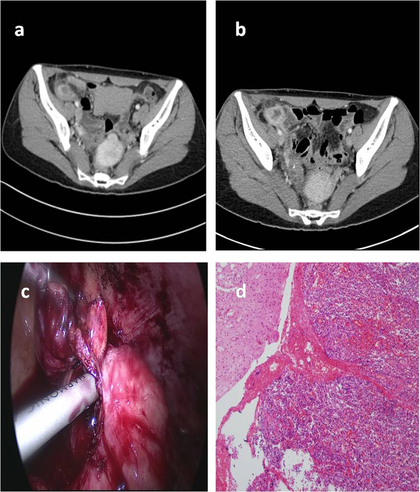
Figure 1. Radiologic, laparoscopic and histologic features of acute appendicitis in an neutropenic patient. Abdominal CT showed wall thickening and edema of the appendix and cecum (a). Abdominal CT showed periappendiceal abscess with increasing periappendiceal and pericecal fat infiltration after relapsed abdominal pain (b). A laparoscopic appendectomy was performed (c), and histologic examination of the appendix revealed infiltration of fibrin and inflammatory cells (hematoxylin-eosin, low magnification × 100) (d).
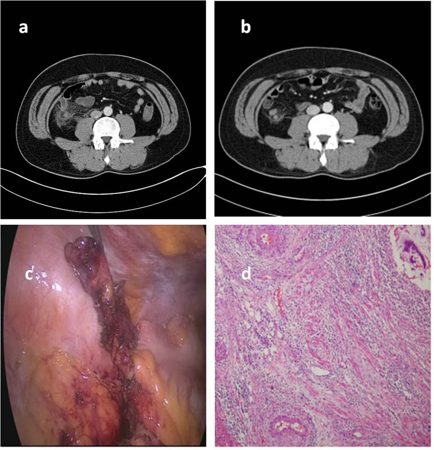
Figure 2. Radiologic, laparoscopic and histologic features of acute appendicitis in an neutropenic leukemic patient. Abdominal CT showed wall thickening and edema of the appendix and periappendiceal inflammatory change (a). Abdominal CT showed an interval decrease of inflammatory process of acute appendicitis with periappendiceal inflammatory change (b). A laparoscopic appendectomy was performed (c), and histologic examination of the appendix revealed infiltration of fibrin and inflammatory cells, without leukemic infiltration (hematoxylin-eosin, low magnification × 100) (d).