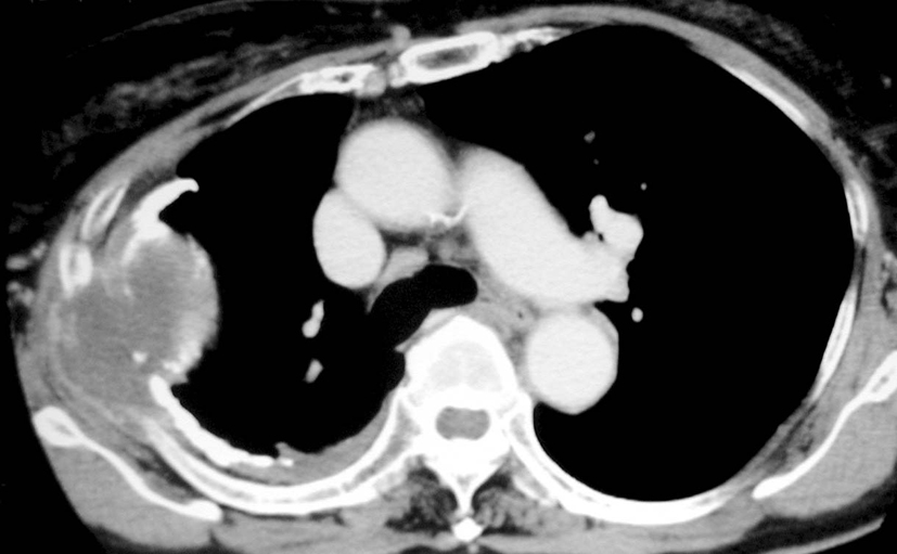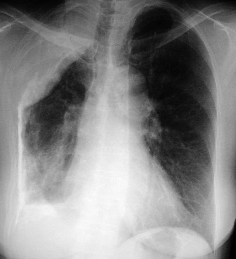
Figure 1. Chest x-ray film on admission shows right-sided pleural calcification and osteolytic changes in the 5th and 6th ribs.
| Journal of Medical Cases, ISSN 1923-4155 print, 1923-4163 online, Open Access |
| Article copyright, the authors; Journal compilation copyright, J Med Cases and Elmer Press Inc |
| Journal website http://www.journalmc.org |
Case Report
Volume 2, Number 2, April 2011, pages 48-50
A Case of Squamous Cell Carcinoma Developed in Chronic Tuberculous Empyema Following Therapeutic Extrapleural Pneumothorax
Figures