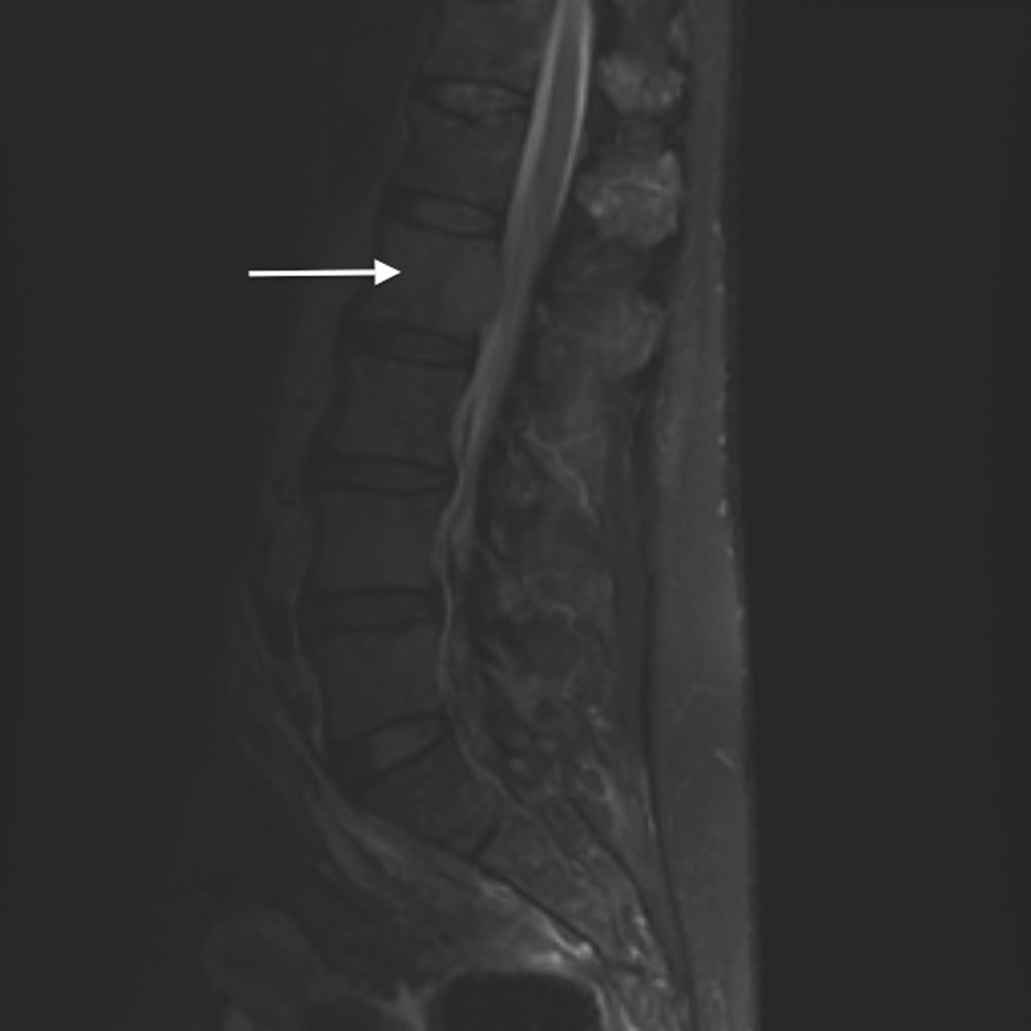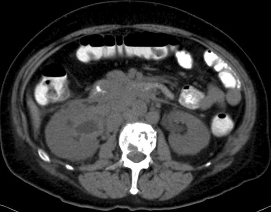
Figure 1. Abdomino-pelvic CT. A diffuse retroperitoneal ganglionnary process with infiltration of the right renal pelvis and the proximal right ureter associated with multiple osseous lytic lesions.
| Journal of Medical Cases, ISSN 1923-4155 print, 1923-4163 online, Open Access |
| Article copyright, the authors; Journal compilation copyright, J Med Cases and Elmer Press Inc |
| Journal website http://www.journalmc.org |
Case Report
Volume 4, Number 7, July 2013, pages 477-480
Obstructive Uropathy by an Extraosseous Multiple Myeloma: A Case Report
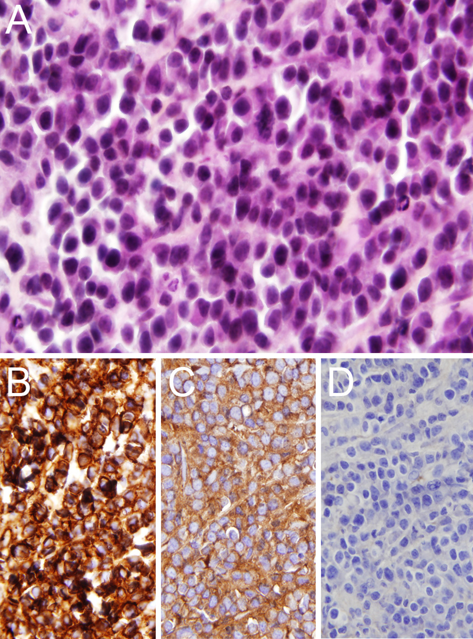
Figures