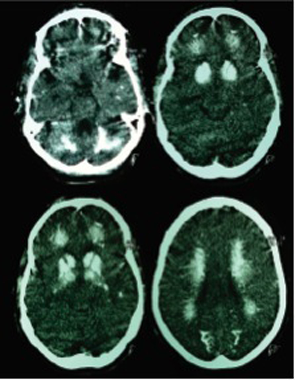
Figure 1. Non-contrast computed tomography scan of the brain showing diffuse calcifications in cerebellum, basal ganglia, semioval centers, thalamus, frontal, temporal, and occipital white matter.
| Journal of Medical Cases, ISSN 1923-4155 print, 1923-4163 online, Open Access |
| Article copyright, the authors; Journal compilation copyright, J Med Cases and Elmer Press Inc |
| Journal website http://www.journalmc.org |
Case Report
Volume 4, Number 6, June 2013, pages 380-384
Fahr’s Syndrome due to Hypoparathyroidism Following Thyroidectomy
Figure
Table
| Classification | Cause |
|---|---|
| Endocrine | Hypoparathyroidsm, pseudohypoparathyroidism pseudopseudohypoparathyroidism, hypothyroidism and hyperparathyroidism. |
| Tumoral | Astrocytoma. |
| Hypoxic and vascular | Ateriovenous malformation, calcified infarct, and ischemic encephalopathy. |
| Inflammatory/Infectious | Cytomegalovirus infection, tuberculosis, measles, toxoplasmosis, neurobrucellosis, mumps, congenital rubella, neurocysticercosis, varicella, acquired immunodeficiency syndrome, coxsackie B infection, and systemic lupus erythematous. |
| Toxic | Carbon monoxide intoxication, lead poisoning, hypervitaminosis D, methotrexate therapy, and radiotherapy. |
| Genetic | Familial idiopathic basal ganglia calcification (Fahr’s disease), idiopathic lenticulodentate calcification (Hastings-James syndrome), Cockayne’s syndrome, Griscelli disease, MELAS (mitochondrial, myopathy, encephalopathy, lactic acidosis, and stroke) syndrome, MERRF (myoclonic epilepsy with ragged red fibers) syndrome, Kearns-Sayre syndrome, Leigh’s disease, Sturge-Weber-Dimitri syndrome, Down’s syndrome, lipoid proteinosis (Urbach-Wieth disease), carbonic anhydrase II deficiency syndrome, biopterin deficit, leukodistrophic diseases, arthrogryposis, and tuberous sclerosis. |
| Other | Senility, malabsorption, motor neuron disease. |