Journal of Medical Cases, ISSN 1923-4155 print, 1923-4163 online, Open Access
Article copyright, the authors; Journal compilation copyright, J Med Cases and Elmer Press Inc
Journal website http://www.journalmc.org

Case Report

Volume 4, Number 5, May 2013, pages 345-348


POEMS Syndrome Manifested as Systemic Capillary Leak Syndrome: A Case Report

Figures

Figure 1.
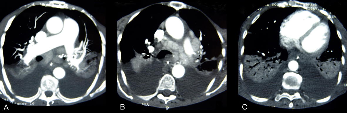
Figure 1. CT pulmonary angiogram had no evidence of pulmonary embolism (A), but disclosed multiple large mediastinal adenomegalies (B) and pleural effusion with atelectasis or consolidation in the adjacent area of the lungs (C).
Figure 2.
Figure 2. Chest RX showing diffuse alveolar filling.
Figure 3.
Figure 3. (Upper) Photomicrograph of mediastinal lymph node stained with HE (original magnification 100 ×): note the reduced germinal center formation and loss of lymph node architecture by mononuclear cell proliferation into interfollicular spaces, as observed in the plasma cell variant of Castleman’s Disease. (Lower) Photomicrograph of mediastinal lymph node showing deposition of hyaline amorphous material and concentric rings of small lymphocytes around vessels, with onion-skin appearance, HE, 400 ×.
Figure 4.
Figure 4. Longitudinal section of trachea showing large lymph nodes on the wall (arrows), in addition to anthracosis (red arrow).