Journal of Medical Cases, ISSN 1923-4155 print, 1923-4163 online, Open Access
Article copyright, the authors; Journal compilation copyright, J Med Cases and Elmer Press Inc
Journal website http://www.journalmc.org

Case Report

Volume 4, Number 6, June 2013, pages 357-361


Differential Diagnosis of Atypical Parathyroid Adenoma and Parathyroid Carcinoma in a Case With Severe Hypercalcemia

Figures

Figure 1.
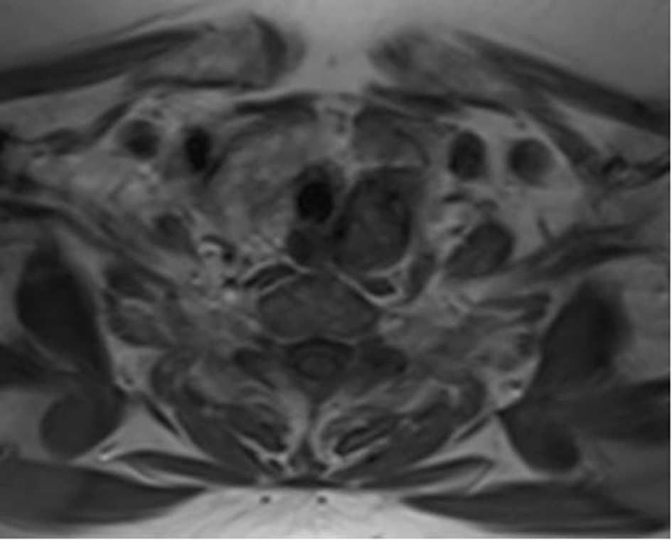
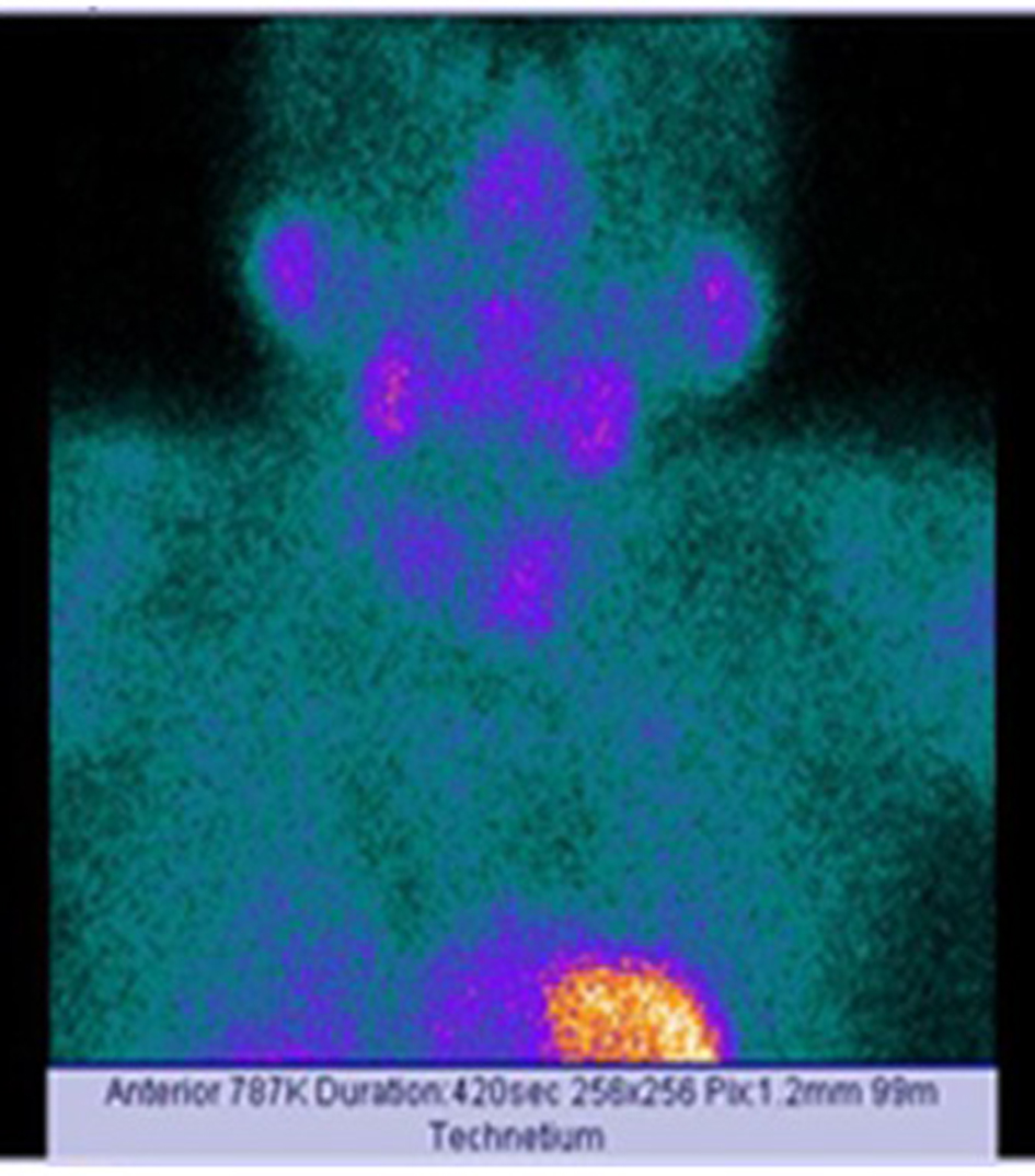
Figure 1. Scanning of the case via Tc-99m sestamibi.
Figure 2.
Figure 2. Nodular lesion at the posterior region of left thyroid lobe on neck magnetic resonance imaging.
Figure 3.
Figure 3. Round to oval shaped proliferative cells seperated by dense fibrotic bands, and cystic and hemorrhagic areas are seen (hematoxyline and eosin stain; original magnification × 50).
Figure 4.
Figure 4. Round to oval shaped parathyroid cell proliferation indicating no marked pleomorphism without mitosis and fat tissue (hematoxyline and eosin stain; original magnification × 400).
Figure 5.
Figure 5. Parathyroid cell proliferation surrounded by thick fibrous capsule without vessel invasion, complete capsule invasion and invasion to the adjacent structures (hematoxyline and eosin stain; original magnification × 50).