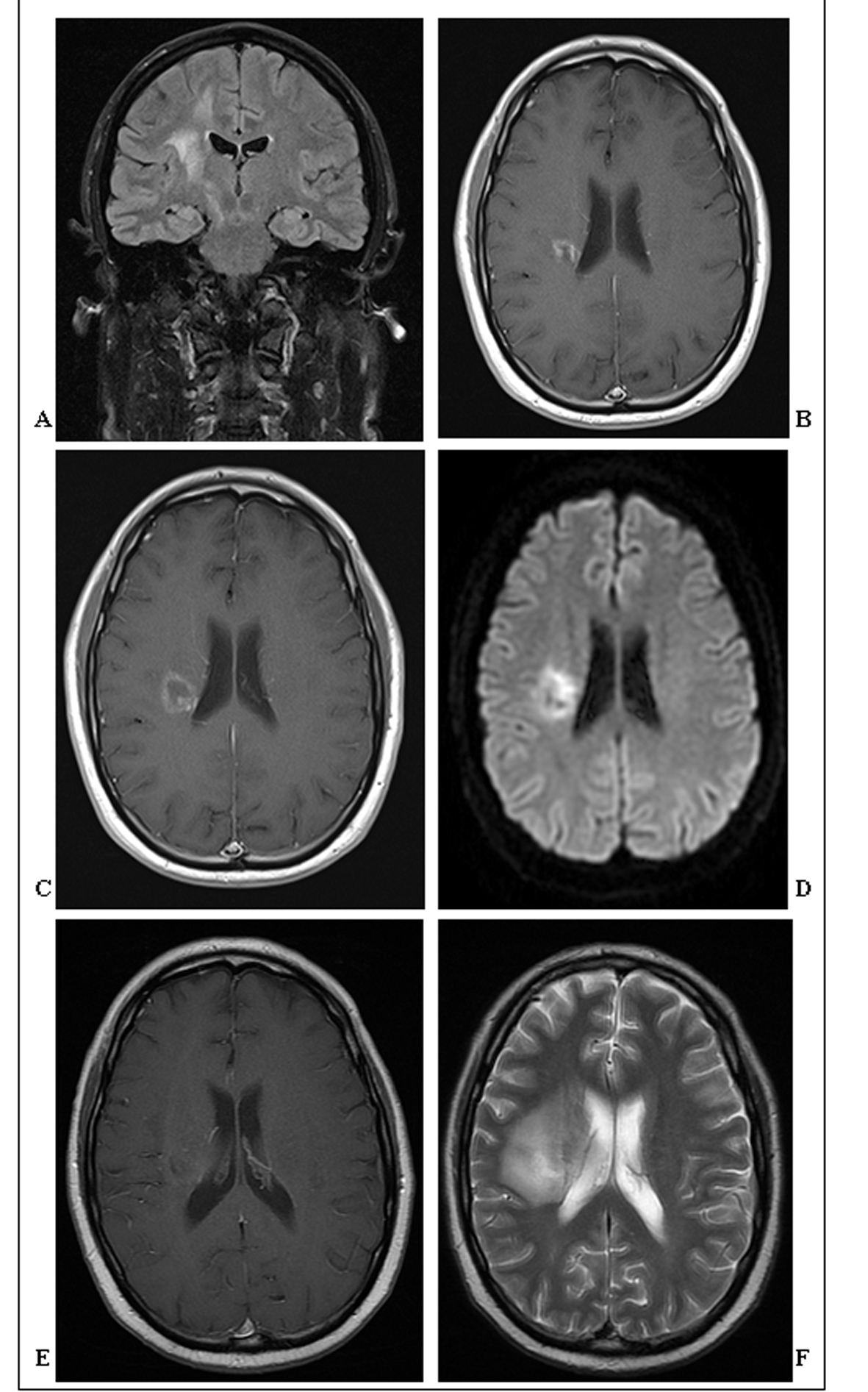
Figure 1. (A). Coronal FLAIR-MRI showing an ill-defined T2 hyperintense 1cm mass in the right posterior centrum semiovale extending inferiorly towards the basal ganglia. Surrounding vasogenic oedema extends throughout adjacent white matter. (B). Axial post contrast T1-weighted MRI showing greater definition of the same enhancing 1.4 cm mass in the right posterior centrum semiovale, now with some central cavitation. (C). Axial post contrast T1-weighted MRI showing further definition, increase in size (2 cm) and central cavitation of the mass in the right posterior centrum semiovale. There is irregular thick ring enhancement of the mass, but no significant mass effect. (D). Axial diffusion-weighted MRI showing restricted diffusion of the centrally cavitating mass in the right posterior centrum semiovale. (E). Axial post-contrast T1-weighted MRI showing poor definition and enhancement of the centrally cavitating mass in the right posterior centrum semiovale. (F). Axial T2-weighted MRI showing the extensive vasogenic oedema surrounding the hyperintense mass in the right posterior centrum semiovale.