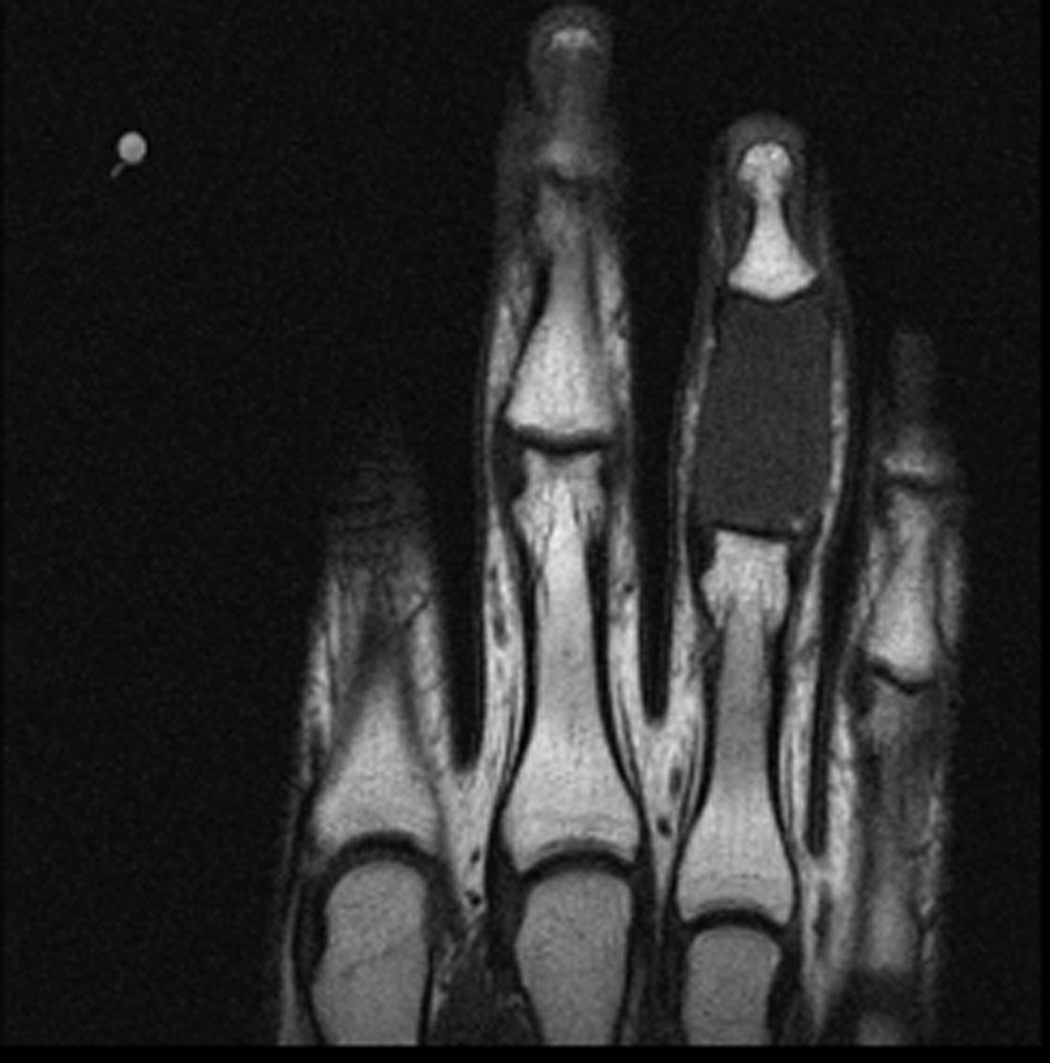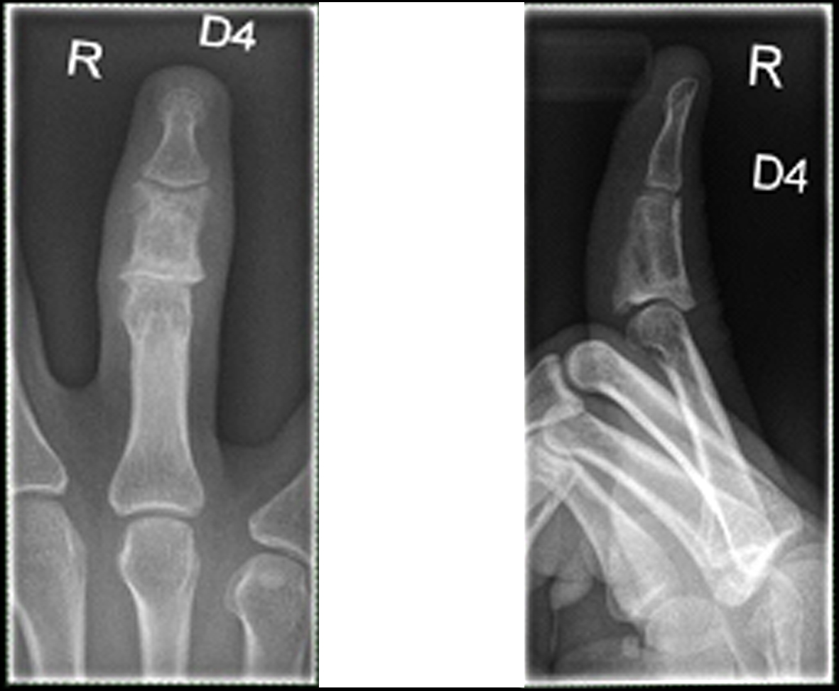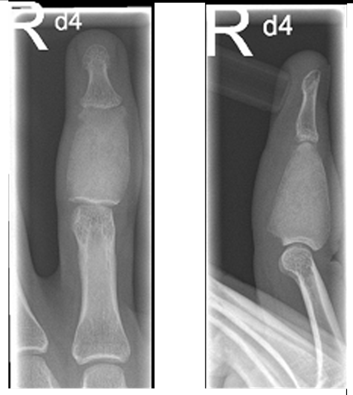
Figure 1. X-ray of the right ring finger: Distended middle phalanx with a ground glass opacity and thinned cortex.
| Journal of Medical Cases, ISSN 1923-4155 print, 1923-4163 online, Open Access |
| Article copyright, the authors; Journal compilation copyright, J Med Cases and Elmer Press Inc |
| Journal website http://www.journalmc.org |
Case Report
Volume 4, Number 5, May 2013, pages 318-322
Monostotic Fibrous Dysplasia of the Middle Phalanx of the Hand
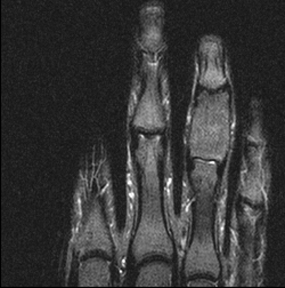
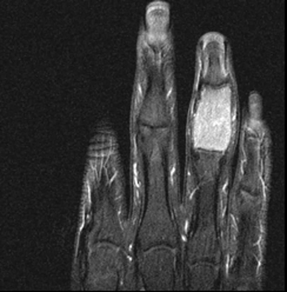
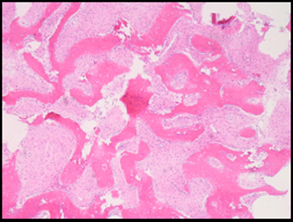
Figures