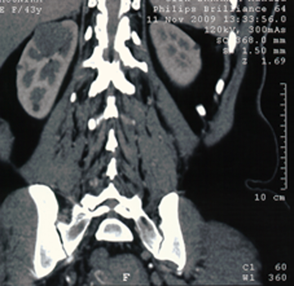
Figure 1. Computed tomography scan: right renal well-defined mass.
| Journal of Medical Cases, ISSN 1923-4155 print, 1923-4163 online, Open Access |
| Article copyright, the authors; Journal compilation copyright, J Med Cases and Elmer Press Inc |
| Journal website http://www.journalmc.org |
Case Report
Volume 1, Number 1, August 2010, pages 18-20
Mucinous Tubular and Spindle Cell Carcinoma of the Kidney: Four New Cases
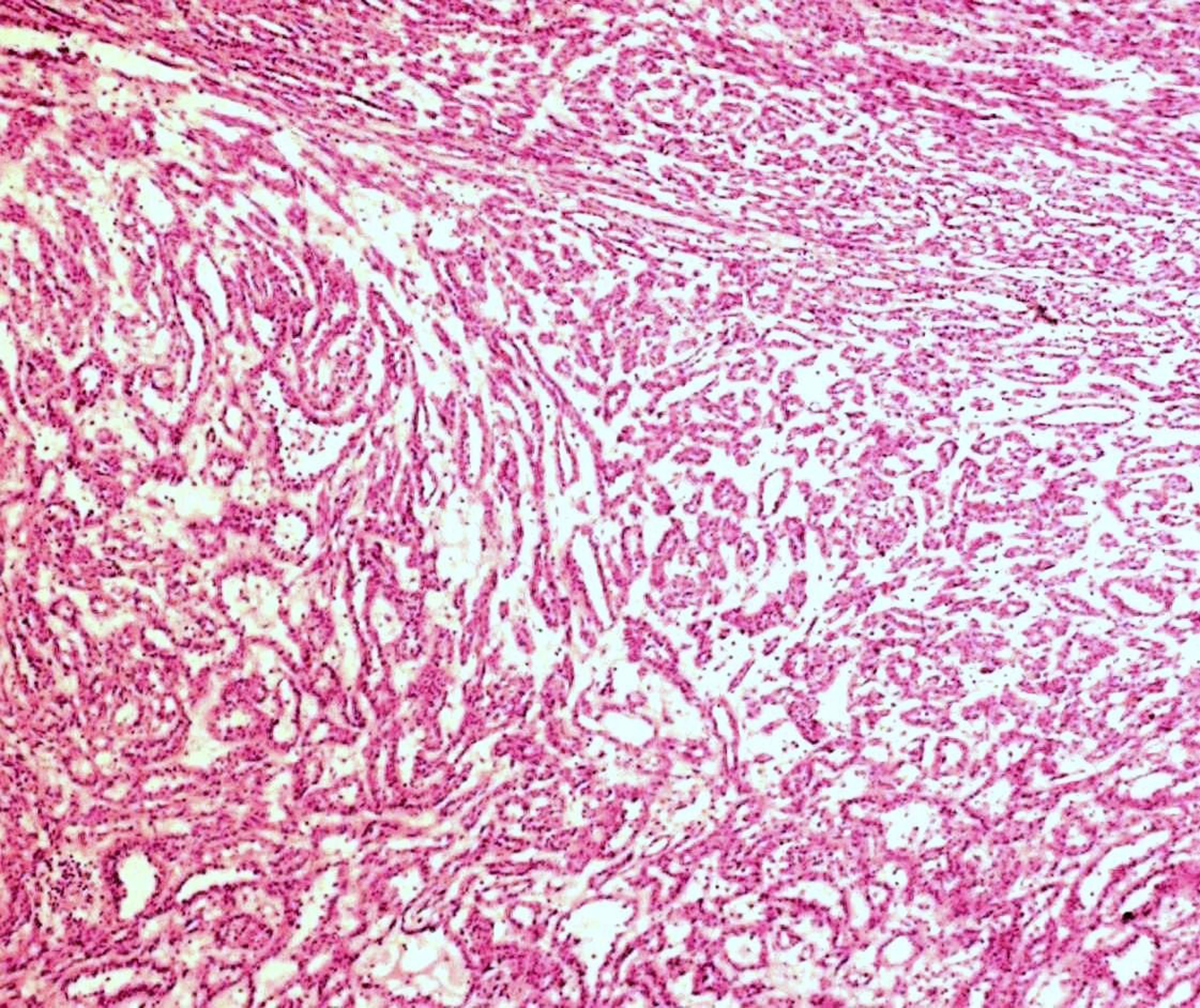
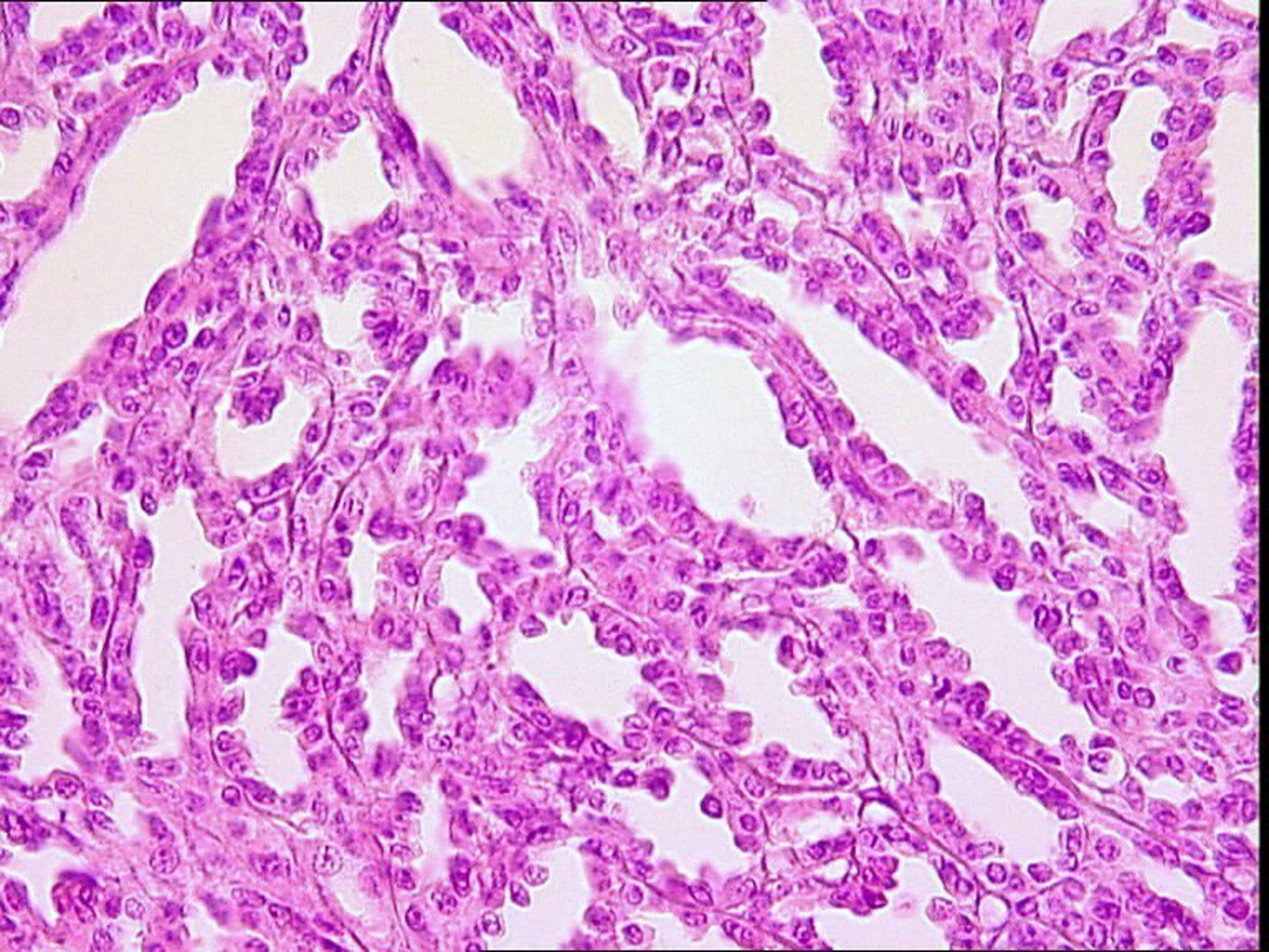
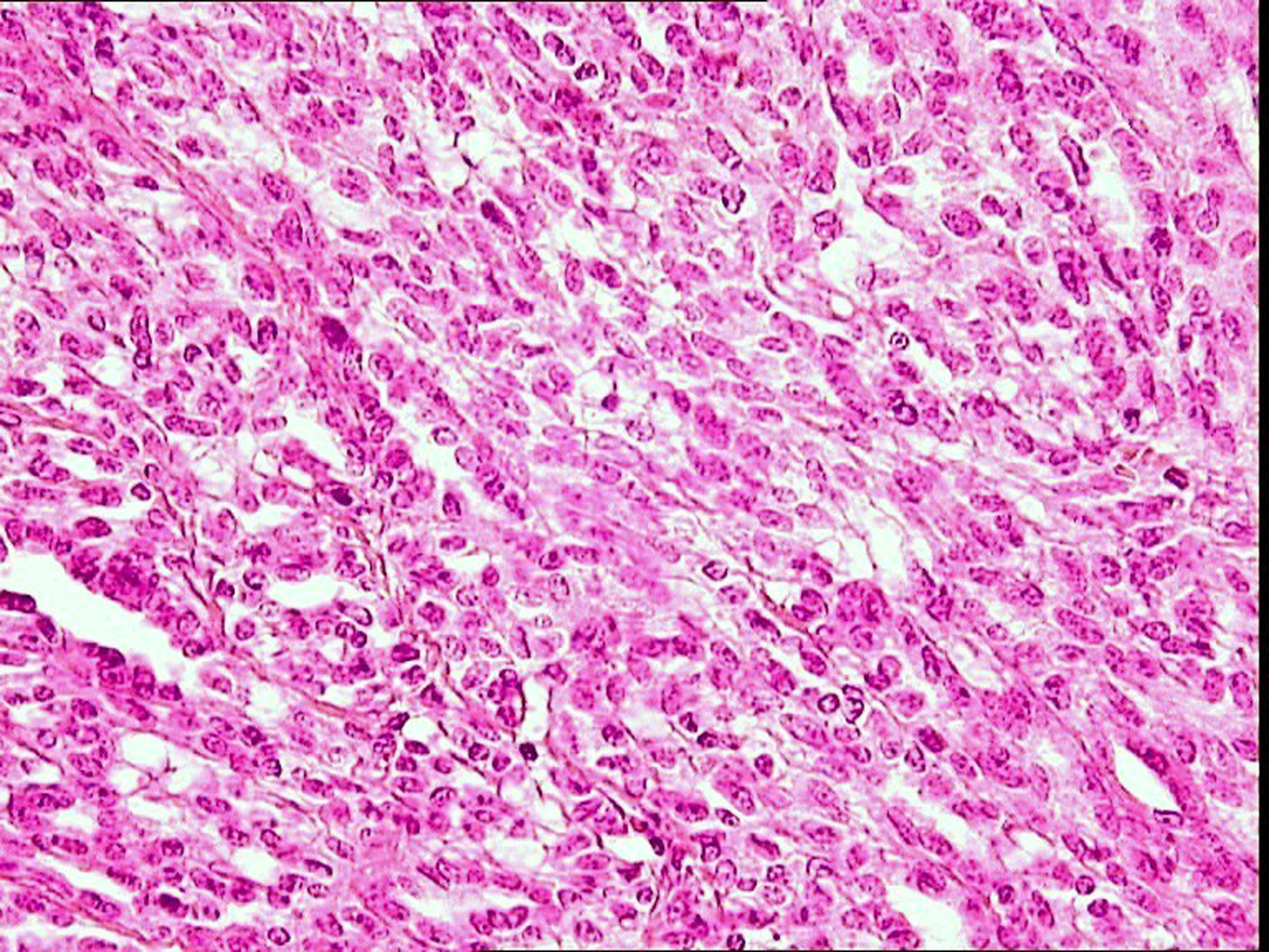
Figures
Table
| Case 1 | Case 2 | Case 3 | Case 4 | |
|---|---|---|---|---|
| Age (years) | 43 | 56 | 45 | 56 |
| Gender | Female | Female | Female | Female |
| Presenting symptoms | Right Asymptomatic | Left Flank pain | Left Flank pain | Left Asymptomatic |
| Tumor size (cm) | 2.5 | 4.5 | 7 | 4 |
| Stage pTNM | pT1N0M0 | pT1N0M0 | pT1N0M0 | pT1N0M0 |
| Treatment | Partial nephrectomy | Radical nephrectomy | Radical nephrectomy | Partial nephrectomy |
| Evolution/follow-up | Favorable 72 months | Favorable 24 months | Favorable 8 months | Favorable 20 months |