Journal of Medical Cases, ISSN 1923-4155 print, 1923-4163 online, Open Access
Article copyright, the authors; Journal compilation copyright, J Med Cases and Elmer Press Inc
Journal website http://www.journalmc.org

Case Report

Volume 1, Number 1, August 2010, pages 1-3


Unusually Severe Bullous Skin Reaction to Sorafenib: A Case Report

Figures

Figure 1.
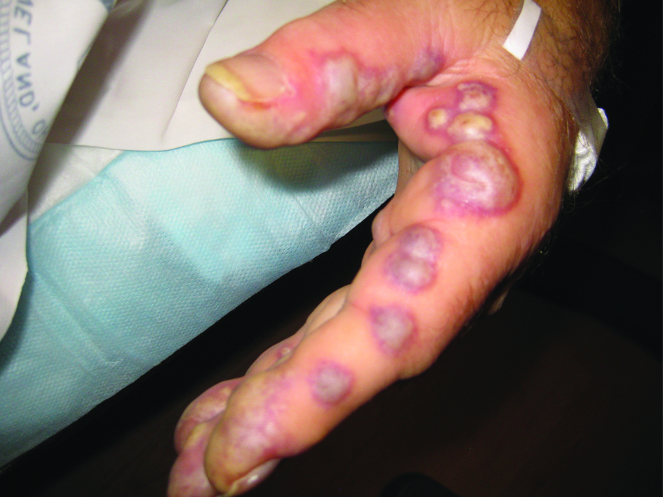
Figure 1. Photo of bullous lesions on patient’s left hand. This picture was taken when both hands had many bullous violaceous lesions. Note the discoloration of the finger tips as well as the bullae along the lateral side of the left index finger. The patient was in such severe pain at this point in his hospital course that he held his hands out in front of him, for fear of the lesions coming into contact with any surface. Later in the hospital course, the lesions erupted, drained, darkened, and became less painful.
Figure 2.
Figure 2. Photomicrograph of acneiform back lesions. This photomicrograph displays the results of the patient’s acneiform back lesion biopsy. Note the ulceration along the top left of the image as well as the neutrophilic infiltration at the bottom. Stained with hematoxylin and eosin, 400x magnification.