Journal of Medical Cases, ISSN 1923-4155 print, 1923-4163 online, Open Access
Article copyright, the authors; Journal compilation copyright, J Med Cases and Elmer Press Inc
Journal website http://www.journalmc.org

Case Report

Volume 4, Number 4, April 2013, pages 247-249


Huge Extrapleural Hematoma Initially Diagnosed as Massive Hemothorax

Figures

Figure 1.
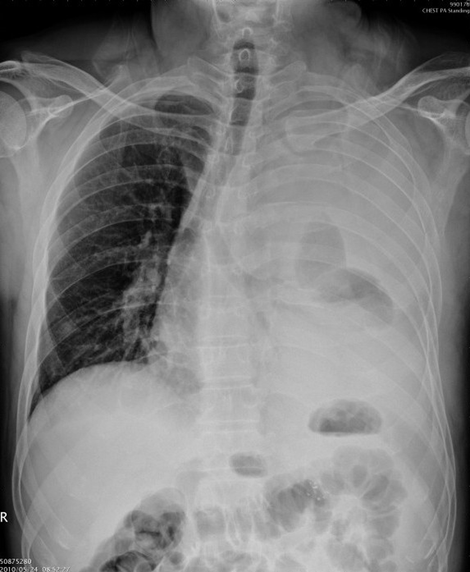
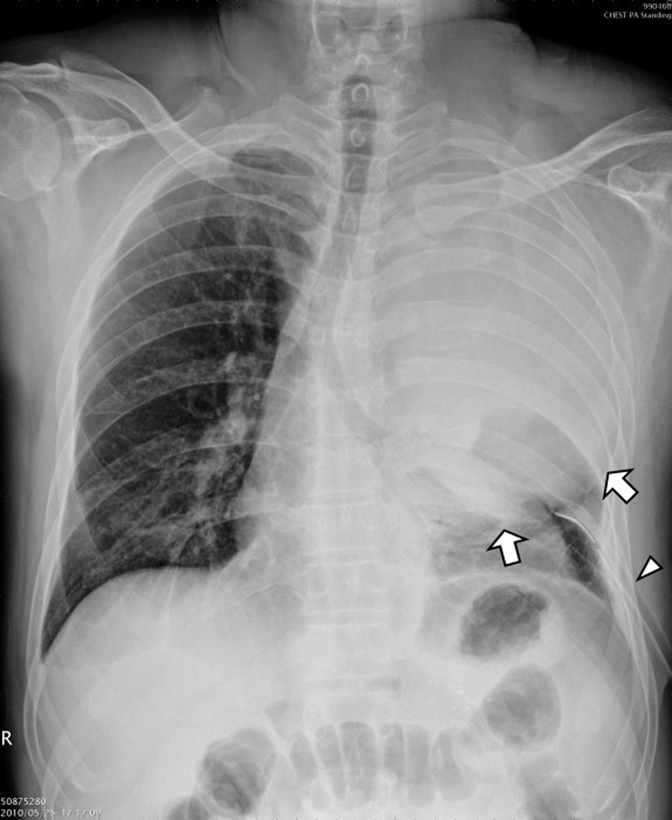
Figure 1. Chest radiograph on admission shows a fracture of the left 4th rib (arrow).
Figure 2.
Figure 2. Complete opacity of the left hemithorax with a shift of the mediastinum to the right 3 days later.
Figure 3.
Figure 3. The left hemithorax still appears as a near complete opacity after insertion of a chest tube (arrowhead). The pleural reflection is identified at the lower margin (arrows) and the costophrenic angle is not obliterated.
Figure 4.
Figure 4. Video-assisted thoracic surgery shows no traumatic pleural collections but a huge pleural mass (arrows).