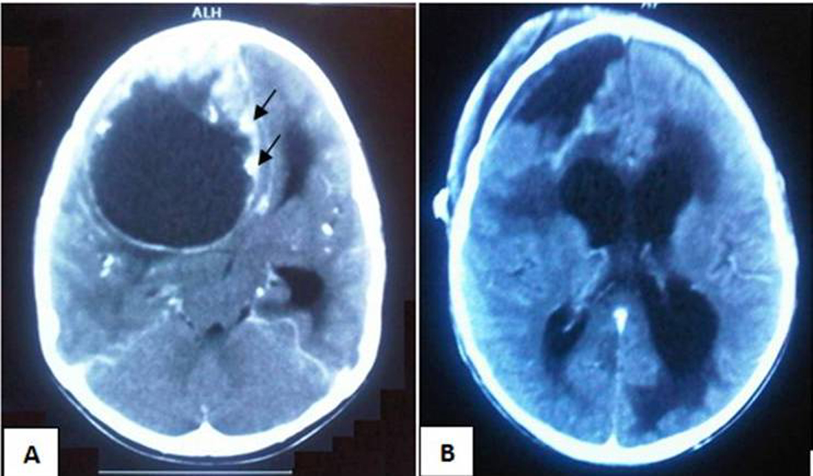
Figure 1. (A): Pre-operative contrasted CT scan depicting huge right frontal lesion including mainly cystic part with solid component along with calcifications (Arrows) and causing obstructive hydrocephalus. (B): Post-operative plain CT scan showing no obvious residual tumor and communicating hydrocephalus.
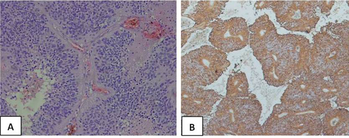
Figure 2. (A): The cytoplasmic processes of ependymal tumor cells condense around blood vessels to form pseudorosettes (Hematoxylin and eosin stain × 400). (B): Glial fibrillary acidic protein (GFAP) highlights cell processes in perivascular pseudorosettes. (GFAP, × 200).
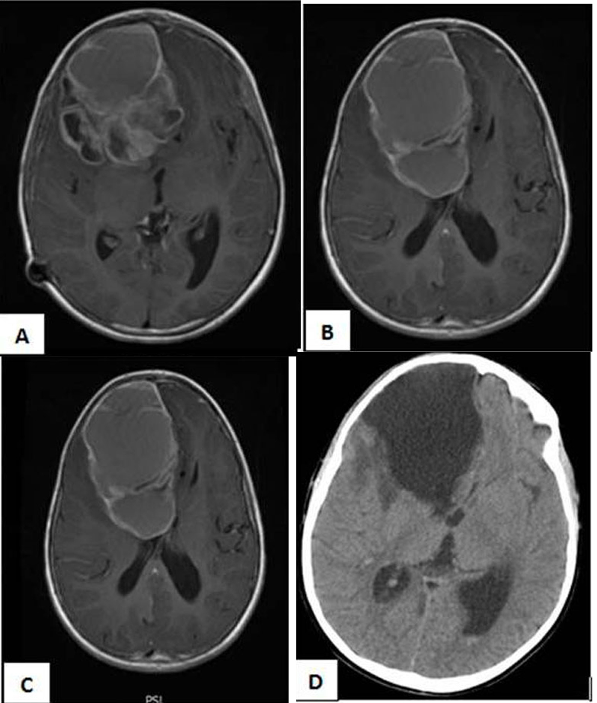
Figure 3. (A, B, C) MRI Brain with Gadolinium, axial cuts showing recurrence of the intracerebral ependymomas with its double enhanced components cystic and solid. (D): Post-operative Ct scan with no residual tumor.