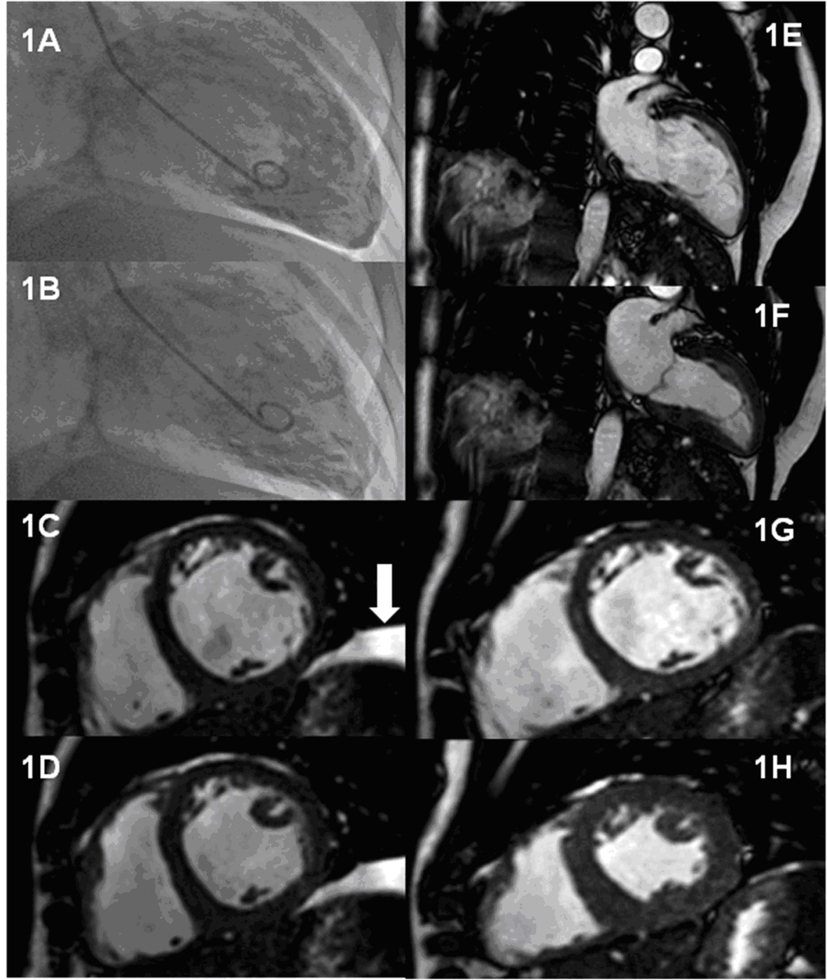
Figure 1. Serial assessment of LV function during treatment with cinacalcet and 4 months after withdrawal. Panels A-B: End-diastolic and end-systolic frames of LV angiogram performed during treatment with cinacalcet following presentation with heart failure symptoms. Panel C-D: Short axis steady state free precession (SSFP) cardiac MR images at the mid ventricular level obtained during treatment Panel C: End-diastolic frame; Panel D: End-systolic frame. The arrow indicates a left sided pleural effusion. Panels E-H: 2 chamber and mid ventricular short axis SSFP cardiac MR images obtained 4 months after discontinuation of cinacalcet. Panels E and G are end-diastolic frames whilst panels F and H are end-systolic frames.