Journal of Medical Cases, ISSN 1923-4155 print, 1923-4163 online, Open Access
Article copyright, the authors; Journal compilation copyright, J Med Cases and Elmer Press Inc
Journal website http://www.journalmc.org

Case Report

Volume 4, Number 2, February 2013, pages 109-113


Duodenal Perforation as an Unusual Celiac Disease Presentation in Two Patients

Figures

Figure 1.
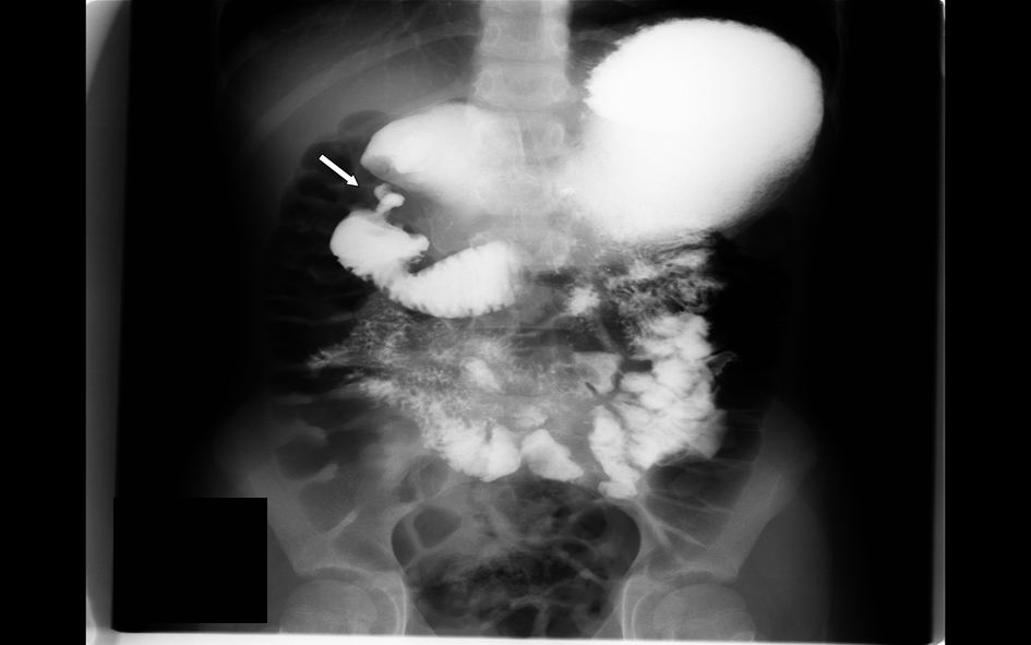
Figure 1. Upper GI barium study showing duodenal bulb dilatation and possible duodenal obstruction.
Figure 2.
Figure 2. CT image showing narrowed descending duodenal lumen with tissue inflammation and thickness around the mid-duodenum suspicious of annular pancreas or intestinal perforation.
Figure 3.
Figure 3. Complete villous atrophy, crypt hyperplasia and intraepithelial lymphocytosis.
Figure 4.
Figure 4. CT image showing free gas and fluid collection in the abdomen posterior to the duodenal bulb with inflammatory changes throughout the mesenteric fat suggestive of intestinal perforation.
Figure 5.
Figure 5. Resected duodenal diverticulum with acute duodenitis with features suggesting celiac disease.